Explore
 Validate
 Learn
Learn Western blot
Antibody data
- Antibody Data
- Antigen structure
- References [0]
- Comments [0]
- Validations
- Western blot [6]
- Immunocytochemistry [1]
- Immunohistochemistry [3]
- Chromatin Immunoprecipitation [1]
Submit
Validation data
Reference
Comment
Report error
- Product number
- PA5-30162 - Provider product page

- Provider
- Invitrogen Antibodies
- Product name
- SATB1 Polyclonal Antibody
- Antibody type
- Polyclonal
- Antigen
- Recombinant protein fragment
- Description
- Recommended positive controls: Jurkat, MDA-MB-231, THP-1, mouse thymus, mouse brain, rat brain.
- Concentration
- 0.78 mg/mL
No comments: Submit comment
Supportive validation
- Submitted by
- Invitrogen Antibodies (provider)
- Main image
- Experimental details
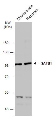
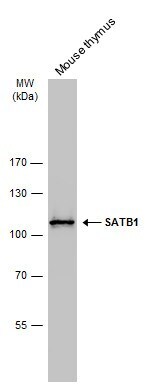
- Western Blot analysis of SATB1 was performed by separating 50 µg of Various tissue extracts by 7.5% SDS-PAGE. Proteins were transferred to a membrane and probed with a SATB1 Polyclonal Antibody (Product # PA5-30162) at a dilution of 1:500. The HRP-conjugated anti-rabbit IgG antibody was used to detect the primary antibody.
- Submitted by
- Invitrogen Antibodies (provider)
- Main image

- Experimental details
- SATB1 Polyclonal Antibody detects SATB1 protein by western blot analysis. Whole cell extracts (30 µg) was separated by 7.5 % SDS-PAGE, and blotted with SATB1 Polyclonal Antibody (Product # PA5-30162) diluted by 1:500.
- Submitted by
- Invitrogen Antibodies (provider)
- Main image
- Experimental details
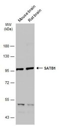
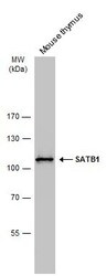
- Western Blot analysis of SATB1 was performed by separating 50 µg of Mouse tissue extracts by 7.5% SDS-PAGE. Proteins were transferred to a membrane and probed with a SATB1 Polyclonal Antibody (Product # PA5-30162) at a dilution of 1:5000. The HRP-conjugated anti-rabbit IgG antibody was used to detect the primary antibody.
- Submitted by
- Invitrogen Antibodies (provider)
- Main image
- Experimental details
- SATB1 Polyclonal Antibody detects SATB1 protein by western blot analysis. Whole cell extracts (30 µg) was separated by 7.5 % SDS-PAGE, and blotted with SATB1 Polyclonal Antibody (Product # PA5-30162) diluted by 1:500.

- Submitted by
- Invitrogen Antibodies (provider)
- Main image
- Experimental details
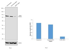
- Knockdown of SATB1 was achieved by transfecting IMR-32 cells with SATB1 specific siRNAs (Silencer® select Product # s12481, Product # s12479). Western blot analysis (Fig. a) was performed using membrane extracts from the SATB1 knockdown cells (Lane 3), non-specific scrambled siRNA transfected cells (Lane 2) and untransfected cells (Lane 1). The blot was probed using Laminin gamma-1 Polyclonal Antibody (Product # PA5-30162, 1:500 dilution) and Goat anti-Rabbit IgG (H+L) Superclonal™ Recombinant Secondary Antibody, HRP (Product # A27036, 1:4000 dilution). Densitometric analysis of this western blot is shown in histogram (Fig. b). Decrease in signal upon siRNA mediated knock down confirms that antibody is specific to SATB1.

- Submitted by
- Invitrogen Antibodies (provider)
- Main image
- Experimental details
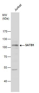
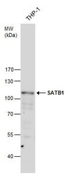
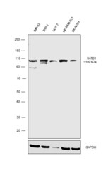
- Western blot was performed using Anti-SATB1 Polyclonal Antibody (Product # PA5-30162) and a ~100 kDa band corresponding to SATB1 was observed across the cell lines tested. Modified whole cell extracts (1% SDS) (30ug lysate) of IMR-32 (Lane 1), THP-1 (Lane 2), MCF-7 (Lane 3), MDA-MB-231 (Lane 4) and SK-N-SH (Lane 5) were electrophoresed using Novex® NuPAGE® 4-12 % Bis-Tris gel (Product # NP0322BOX). Resolved proteins were then transferred onto a nitrocellulose membrane (Product # IB23001) by iBlot® 2 Dry Blotting System (Product # IB21001). The blot was probed with the primary antibody (1:500 dilution) and detected by chemiluminescence with Goat anti-Rabbit IgG (H+L) Superclonal™ Recombinant Secondary Antibody, HRP (Product # A27036, 1:4000 dilution) using the iBright FL 1000 (Product # A32752). Chemiluminescent detection was performed using Novex® ECL Chemiluminescent Substrate Reagent Kit (Product # WP20005).

Supportive validation
- Submitted by
- Invitrogen Antibodies (provider)
- Main image

- Experimental details
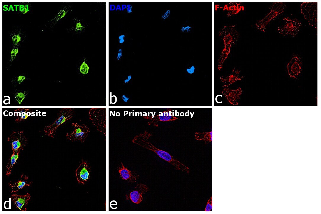
- Immunofluorescence analysis of SATB1 was performed using 70% confluent log phase MDA-MB-231 cells. The cells were fixed with 4% Paraformaldehyde for 10 minutes, permeabilized with 0.1% Triton™ X-100 for 15 minutes, and blocked with 2% BSA for 10 minutes at room temperature. The cells were labeled with SATB1 Polyclonal Antibody (Product # PA5-30162) at 1:500 dilution in 0.1% BSA, incubated at 4 degree Celsius overnight and then labeled with Goat anti-Rabbit IgG (H+L) Superclonal™ Recombinant Secondary Antibody (Product # A27034) at a dilution of 1:2000 for 45 minutes at room temperature (Panel a: Green). Nuclei (Panel b: Blue) were stained with DAPI. F-actin (Panel c: Red) was stained with Rhodamine Phalloidin (Product # R415, 1:300). Panel d represents the merged image showing Nuclear and Cytoplasmic localization. Panel e represents control cells with no primary antibody to assess background. The images were captured at 60X magnification.
Supportive validation
- Submitted by
- Invitrogen Antibodies (provider)
- Main image
- Experimental details
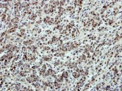
- Immunohistochemical analysis of paraffin-embedded TOV-21G xenograft, using SATB1 (Product # PA5-30162) antibody at 1:500 dilution. Antigen Retrieval: EDTA based buffer, pH 8.0, 15 min.
- Submitted by
- Invitrogen Antibodies (provider)
- Main image
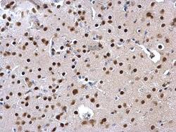
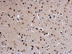
- Experimental details
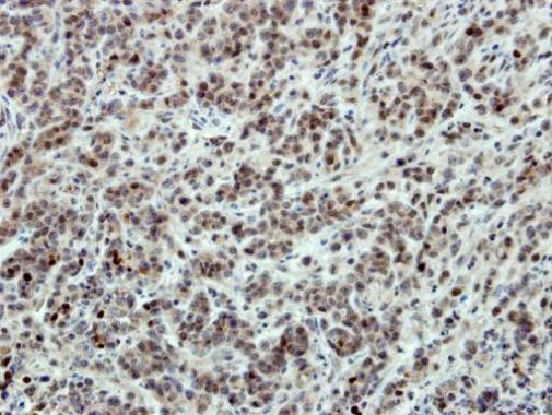
- SATB1 Polyclonal Antibody detects SATB1 protein at nucleus on mouse fore brain by immunohistochemical analysis. Sample: Paraffin-embedded mouse fore brain. SATB1 Polyclonal Antibody (Product # PA5-30162) dilution: 1:500. Antigen Retrieval: EDTA based buffer, pH 8.0, 15 min.
- Submitted by
- Invitrogen Antibodies (provider)
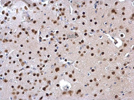
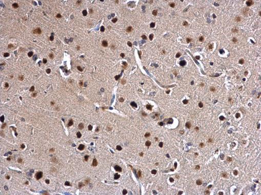
- Main image
- Experimental details
- SATB1 Polyclonal Antibody detects SATB1 protein at nucleus on rat middle brain by immunohistochemical analysis. Sample: Paraffin-embedded rat middle brain. SATB1 Polyclonal Antibody (Product # PA5-30162) dilution: 1:500. Antigen Retrieval: EDTA based buffer, pH 8.0, 15 min.
Supportive validation
- Submitted by
- Invitrogen Antibodies (provider)
- Main image
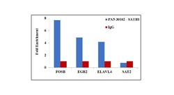
- Experimental details
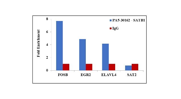
- Chromatin Immunoprecipitation (ChIP) assay of endogenous SATB1 protein using Anti-SATB1 Antibody: ChIP was performed using Anti-SATB1 Rabbit polyclonal Antibody (Product # PA5-30162, 5 µg) on sheared chromatin from IMR32 cells using the MAGnify ChIP System kit (Product # 49-2024). Normal Rabbit IgG was used as a negative IP control. The purified DNA was analyzed by qPCR using primers binding to FOSB transcriptional start site, EGR2 intronic region (+2.5kb), ELAVL4 exonic region (+59kb) and SAT2 satellite repeats. Data is presented as fold enrichment of the antibody signal versus the negative control IgG using the comparative CT method.