 Explore
 Validate
 Learn
Learn Western blot
 Immunocytochemistry
Antibody data
- Antibody Data
- Antigen structure
- References [1]
- Comments [0]
- Validations
- Immunocytochemistry [2]
- Immunoprecipitation [1]
- Immunohistochemistry [3]
- Chromatin Immunoprecipitation [2]
- Other assay [2]
Submit
Validation data
Reference
Comment
Report error
- Product number
- PA5-30163 - Provider product page

- Provider
- Invitrogen Antibodies
- Product name
- SATB1 Polyclonal Antibody
- Antibody type
- Polyclonal
- Antigen
- Recombinant full-length protein
- Description
- Recommended positive controls: Jurkat, THP-1, mouse thymus. Predicted reactivity: Mouse (96%), Rat (96%), Chicken (91%), Rhesus Monkey (99%). Store product as a concentrated solution. Centrifuge briefly prior to opening the vial.
- Reactivity
- Human, Mouse
- Host
- Rabbit
- Isotype
- IgG
- Vial size
- 100 μL
- Concentration
- 0.36 mg/mL
- Storage
- Store at 4°C short term. For long term storage, store at -20°C, avoiding freeze/thaw cycles.
Submitted references Inhibition of prostate cancer DU145 cell growth with small interfering RNA targeting the SATB1 gene.
Wang Q, Yang CS, Ma ZX, Chen JC, Zheng JN, Sun XQ, Wang JQ
Experimental and therapeutic medicine 2018 Mar;15(3):3028-3033
Experimental and therapeutic medicine 2018 Mar;15(3):3028-3033
No comments: Submit comment
Supportive validation
- Submitted by
- Invitrogen Antibodies (provider)
- Main image

- Experimental details
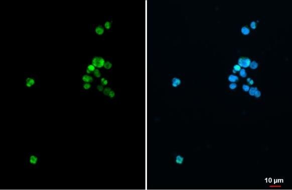
- SATB1 Polyclonal Antibody detects SATB1 protein at nucleus by immunofluorescent analysis. Sample: Jurkat cells were fixed in 4% paraformaldehyde at RT for 15 min. Green: SATB1 stained by SATB1 Polyclonal Antibody (Product # PA5-30163) diluted at 1:500. Blue: Fluoroshield with DAPI .

- Submitted by
- Invitrogen Antibodies (provider)
- Main image

- Experimental details
- SATB1 Polyclonal Antibody detects SATB1 protein at nucleus by immunofluorescent analysis. Sample: Jurkat cells were fixed in 4% paraformaldehyde at RT for 15 min. Green: SATB1 stained by SATB1 Polyclonal Antibody (Product # PA5-30163) diluted at 1:500. Blue: Fluoroshield with DAPI .
Supportive validation
- Submitted by
- Invitrogen Antibodies (provider)
- Main image
- Experimental details
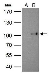
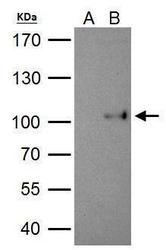
- SATB1 antibody immunoprecipitates SATB1 protein in IP experiments. IP Sample: 293T whole cell lysate/extract A. Control with 2 µg of preimmune rabbit IgG B. Immunoprecipitation of SATB1 protein by 2 µg of SATB1 antibody (Product # PA5-30163) 7.5% SDS-PAGE The immunoprecipitated SATB1 protein was detected by SATB1 antibody (Product # PA5-30163) diluted at 1:1,000.

Supportive validation
- Submitted by
- Invitrogen Antibodies (provider)
- Main image
- Experimental details
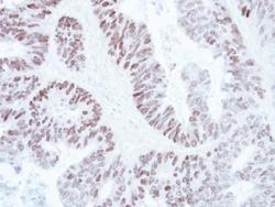
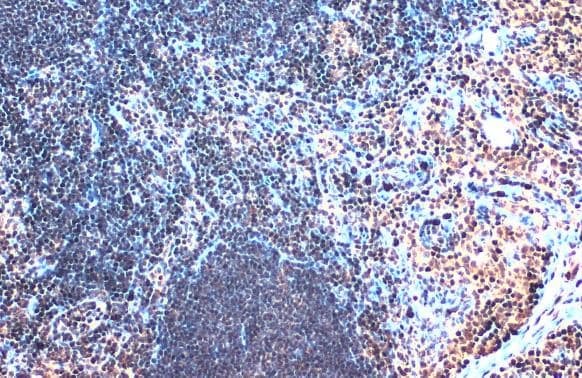
- Immunohistochemical analysis of paraffin-embedded human colon carcinoma, using SATB1 (Product # PA5-30163) antibody at 1:250 dilution. Antigen Retrieval: EDTA based buffer, pH 8.0, 15 min.
- Submitted by
- Invitrogen Antibodies (provider)
- Main image
- Experimental details
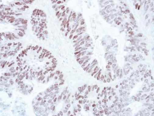
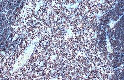
- SATB1 Polyclonal Antibody detects SATB1 protein at nucleus by immunohistochemical analysis. Sample: Paraffin-embedded mouse thymus gland. SATB1 stained by SATB1 Polyclonal Antibody (Product # PA5-30163) diluted at 1:500. Antigen Retrieval: Citra.

- Submitted by
- Invitrogen Antibodies (provider)
- Main image

- Experimental details
- SATB1 Polyclonal Antibody detects SATB1 protein at nucleus by immunohistochemical analysis. Sample: Paraffin-embedded mouse lymph node. SATB1 stained by SATB1 Polyclonal Antibody (Product # PA5-30163) diluted at 1:500. Antigen Retrieval: Citrate.
Supportive validation
- Submitted by
- Invitrogen Antibodies (provider)
- Main image

- Experimental details
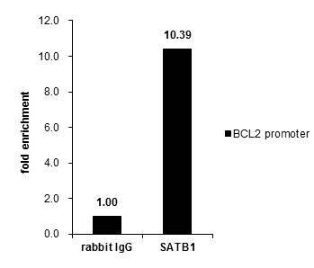
- Cross-linked ChIP was performed with Jurkat chromatin extract and 5 µg of either control rabbit IgG or SATB1 Polyclonal Antibody (Product # PA5-30163). The precipitated DNA was detected by PCR with primer set targeting to BCL2 promoter.

- Submitted by
- Invitrogen Antibodies (provider)
- Main image

- Experimental details
- Cross-linked ChIP was performed with Jurkat chromatin extract and 5 µg of either control rabbit IgG or SATB1 Polyclonal Antibody (Product # PA5-30163). The precipitated DNA was detected by PCR with primer set targeting to BCL2 promoter.
Supportive validation
- Submitted by
- Invitrogen Antibodies (provider)
- Main image
- Experimental details
- SATB1 antibody immunoprecipitates SATB1 protein in IP experiments. IP Sample: 293T whole cell lysate/extract A. Control with 2 µg of preimmune rabbit IgG B. Immunoprecipitation of SATB1 protein by 2 µg of SATB1 antibody (Product # PA5-30163) 7.5% SDS-PAGE The immunoprecipitated SATB1 protein was detected by SATB1 antibody (Product # PA5-30163) diluted at 1:1,000.
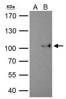
- Submitted by
- Invitrogen Antibodies (provider)
- Main image
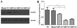
- Experimental details
- Figure 3. Protein expression levels of SATB1. (A) Western blot analysis was performed to determine SATB1 protein expression levels following siRNA2 transfection compared with the NC and Blank groups (n=5). (B) ImageJ quantitation of the band densities from the western blot analysis of SATB1 was performed. *P
