 Explore
 Validate
 Learn
Learn Western blot
 Immunoprecipitation
Antibody data
- Antibody Data
- Antigen structure
- References [0]
- Comments [0]
- Validations
- Western blot [3]
- Immunocytochemistry [2]
- Immunohistochemistry [4]
- Other assay [1]
Submit
Validation data
Reference
Comment
Report error
- Product number
- 60265-1-IG - Provider product page

- Provider
- Invitrogen Antibodies
- Product name
- SND1 Monoclonal Antibody (1A6A4)
- Antibody type
- Monoclonal
- Antigen
- Other
- Reactivity
- Human, Mouse, Rat
- Host
- Mouse
- Isotype
- IgG
- Antibody clone number
- 1A6A4
- Vial size
- 150 µL
- Concentration
- 1 mg/mL
- Storage
- -20°C
No comments: Submit comment
Supportive validation
- Submitted by
- Invitrogen Antibodies (provider)
- Main image
- Experimental details
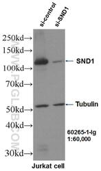
- WB result of SND1 antibody (60265-1-IG, 1:60,000) with si-Control and si-SND1 transfected Jurkat cells.
- Submitted by
- Invitrogen Antibodies (provider)
- Main image
- Experimental details
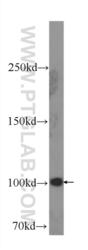
- HeLa cells were subjected to SDS PAGE followed by western blot with 60265-1-IG ( SND1 Antibody) at dilution of 1:1000 incubated at room temperature for 1.5 hours.

- Submitted by
- Invitrogen Antibodies (provider)
- Main image

- Experimental details
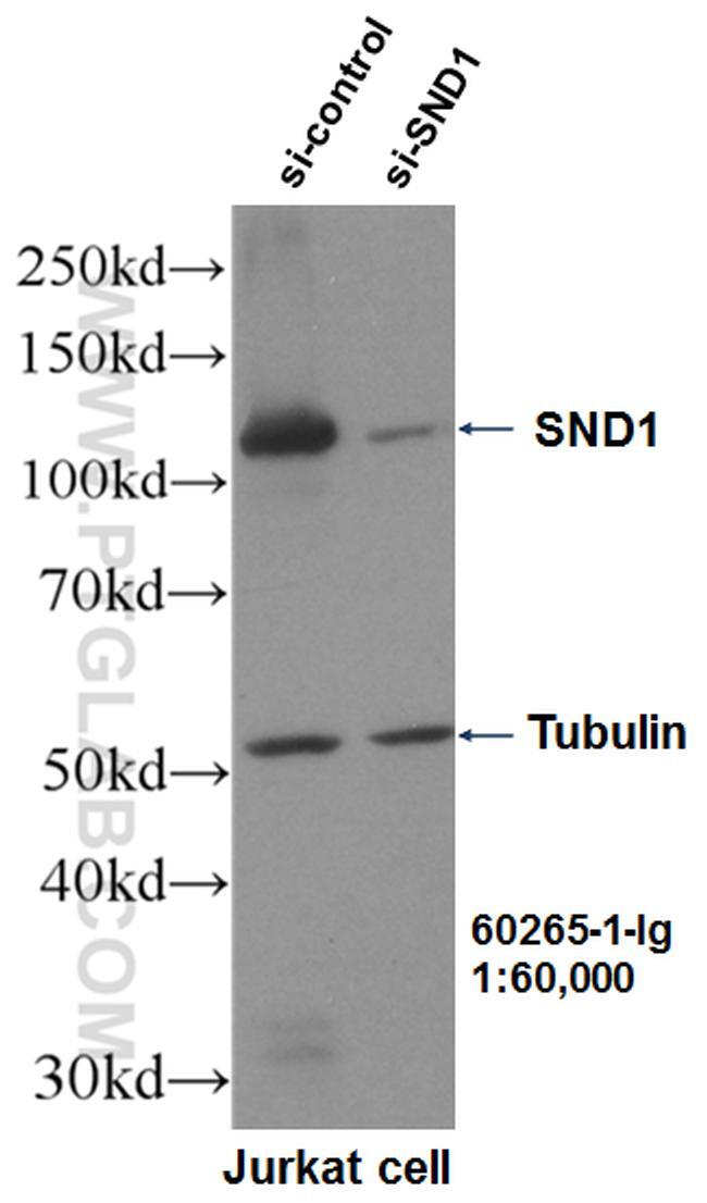
- Jurkat cells were subjected to SDS PAGE followed by western blot with 60265-1-Ig (SND1 antibody) at dilution of 1:1000 incubated at room temperature for 1.5 hours.
Supportive validation
- Submitted by
- Invitrogen Antibodies (provider)
- Main image
- Experimental details
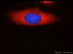
- Immunofluorescent analysis of HepG2 cells using 60265-1-IG (SND1 antibody) at dilution of 1:50 and and Rhodamine-labeled goat anti-mouse IGG (red).
- Submitted by
- Invitrogen Antibodies (provider)
- Main image
- Experimental details
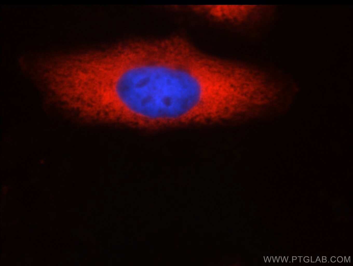
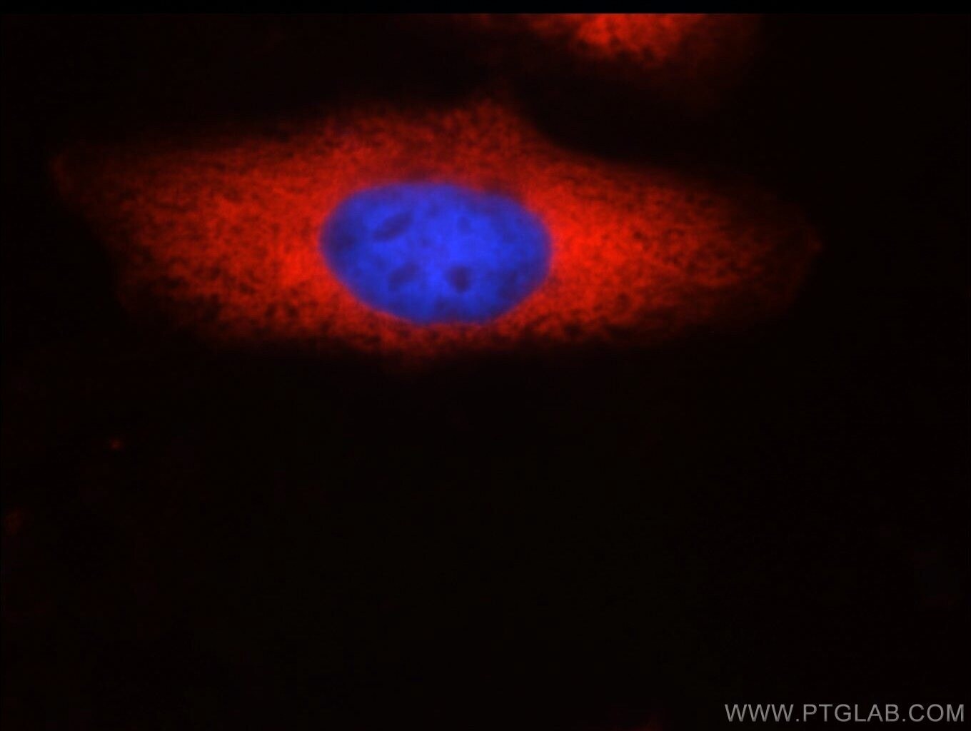
- Immunofluorescent analysis of HepG2 cells using 60265-1-IG (SND1 antibody) at dilution of 1:50 and and Rhodamine-labeled goat anti-mouse IGG (red).
Supportive validation
- Submitted by
- Invitrogen Antibodies (provider)
- Main image
- Experimental details
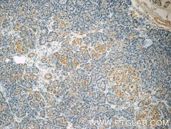
- Immunohistochemistry of paraffin-embedded human pancreas tissue slide using 60265-1-Ig (SND1 Antibody) at dilution of 1:50 (under 10x lens).
- Submitted by
- Invitrogen Antibodies (provider)
- Main image

- Experimental details
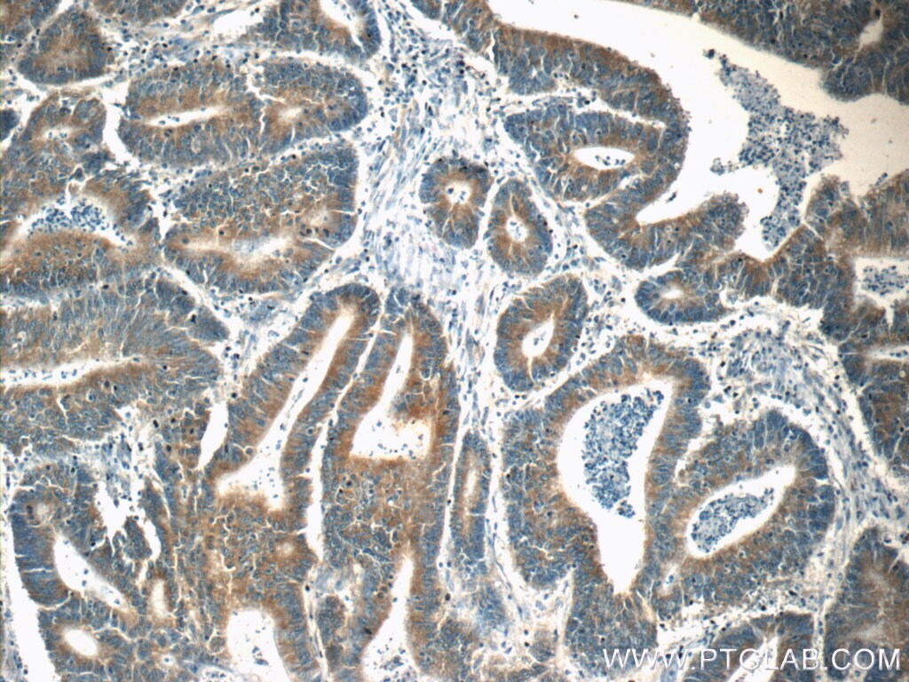
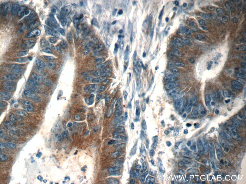
- Immunohistochemistry of paraffin-embedded human colon cancer tissue slide using 60265-1-Ig (SND1 Antibody) at dilution of 1:50 (under 10x lens).
- Submitted by
- Invitrogen Antibodies (provider)
- Main image
- Experimental details
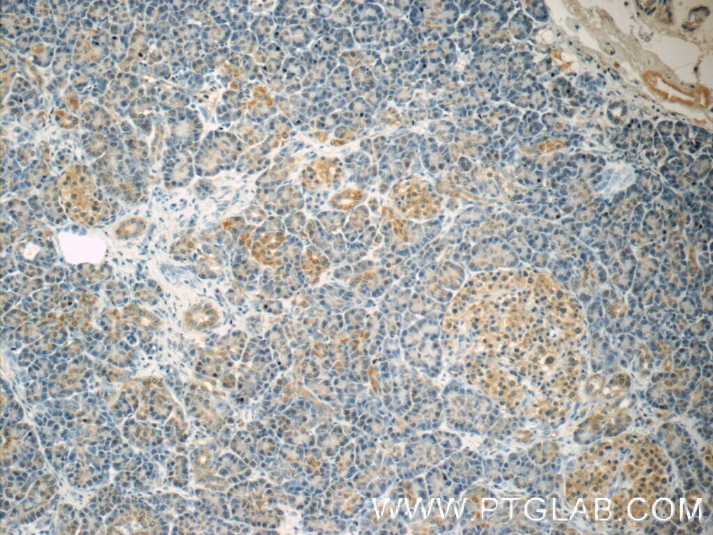
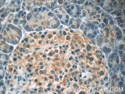
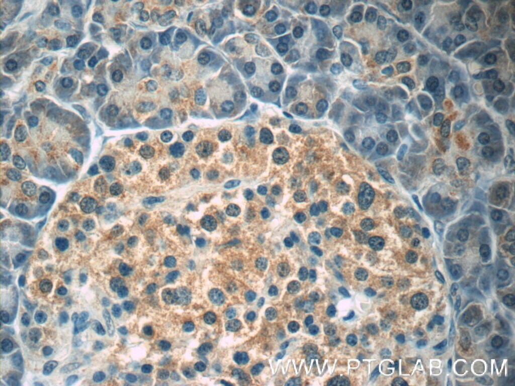
- Immunohistochemistry of paraffin-embedded human pancreas tissue slide using 60265-1-IG ( SND1 Antibody) at dilution of 1:50 (under 40x lens).
- Submitted by
- Invitrogen Antibodies (provider)
- Main image

- Experimental details
- Immunohistochemistry of paraffin-embedded human colon cancer tissue slide using 60265-1-IG ( SND1 Antibody) at dilution of 1:50 (under 40x lens).
Supportive validation
- Submitted by
- Invitrogen Antibodies (provider)
- Main image
- Experimental details
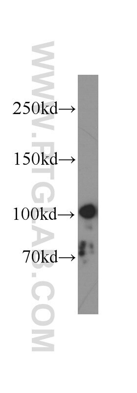
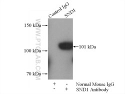
- IP result of anti-SND1 (IP:60265-1-IG, 5ug; Detection:60265-1-IG 1:500) with HeLa cells lysate 1400ug.
