Explore
 Validate
 Learn
Learn Western blot
 Immunohistochemistry
Antibody data
- Antibody Data
- Antigen structure
- References [1]
- Comments [0]
- Validations
- Immunohistochemistry [1]
Submit
Validation data
Reference
Comment
Report error
- Product number
- HPA002529 - Provider product page

- Provider
- Atlas Antibodies
- Proper citation
- Atlas Antibodies Cat#HPA002529, RRID:AB_1857317
- Product name
- Anti-SND1
- Antibody type
- Polyclonal
- Description
- Polyclonal Antibody against Human SND1, Gene description: staphylococcal nuclease and tudor domain containing 1, Alternative Gene Names: p100, TDRD11, Validated applications: WB, IHC, Uniprot ID: Q7KZF4, Storage: Store at +4°C for short term storage. Long time storage is recommended at -20°C.
- Reactivity
- Human, Mouse, Rat
- Host
- Rabbit
- Conjugate
- Unconjugated
- Isotype
- IgG
- Vial size
- 100 µl
- Concentration
- 0.1 mg/ml
- Storage
- Store at +4°C for short term storage. Long time storage is recommended at -20°C.
- Handling
- The antibody solution should be gently mixed before use.
Submitted references Pharmacological disruption of the MTDH–SND1 complex enhances tumor antigen presentation and synergizes with anti-PD-1 therapy in metastatic breast cancer
Shen M, Smith H, Wei Y, Jiang Y, Zhao S, Wang N, Rowicki M, Tang Y, Hang X, Wu S, Wan L, Shao Z, Kang Y
Nature Cancer 2021;3(1):60-74
Nature Cancer 2021;3(1):60-74
No comments: Submit comment
Supportive validation
- Submitted by
- Atlas Antibodies (provider)
- Enhanced method
- Orthogonal validation
- Main image

- Experimental details
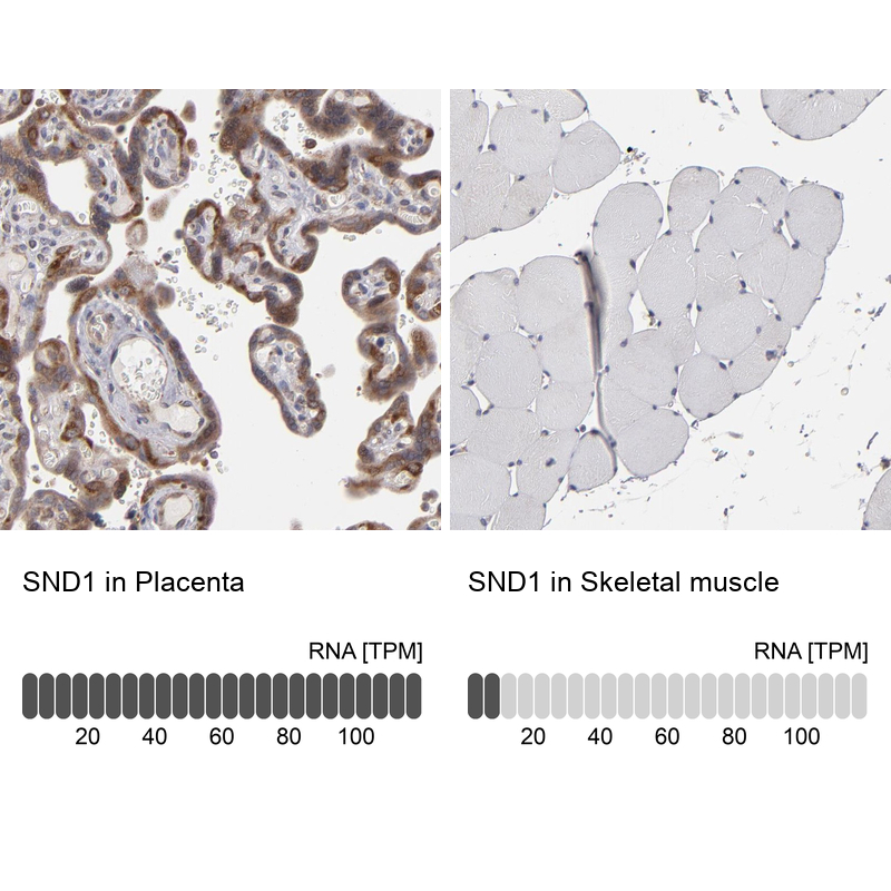
- Immunohistochemistry analysis in human placenta and skeletal muscle tissues using Anti-SND1 antibody. Corresponding SND1 RNA-seq data are presented for the same tissues.
- Sample type
- Human
- Protocol
- Protocol