Explore
 Validate
 Learn
LearnPA5-54425
antibody from Invitrogen Antibodies
Targeting: WIPI2
ATG18B, ATG21, CGI-50, DKFZP434J154, DKFZp686P02188, FLJ12979, FLJ14217, FLJ42984
 Western blot
 Immunocytochemistry
Antibody data
- Antibody Data
- Antigen structure
- References [0]
- Comments [0]
- Validations
- Immunocytochemistry [1]
Submit
Validation data
Reference
Comment
Report error
- Product number
- PA5-54425 - Provider product page

- Provider
- Invitrogen Antibodies
- Product name
- WIPI2 Polyclonal Antibody
- Antibody type
- Polyclonal
- Antigen
- Recombinant full-length protein
- Description
- Immunogen sequence: MKVLHTIRET PPNPAGLCAL SINNDNCYLA YPGSATIGEV QVFDTINLRA ANMIPAHDSP LAALAFDASG TKLATASEKG TVIRVF Highest antigen sequence identity to the following orthologs: Mouse - 99%, Rat - 100%.
- Reactivity
- Human
- Host
- Rabbit
- Isotype
- IgG
- Vial size
- 100 µL
- Concentration
- 0.2 mg/mL
- Storage
- Store at 4°C short term. For long term storage, store at -20°C, avoiding freeze/thaw cycles.
No comments: Submit comment
Supportive validation
- Submitted by
- Invitrogen Antibodies (provider)
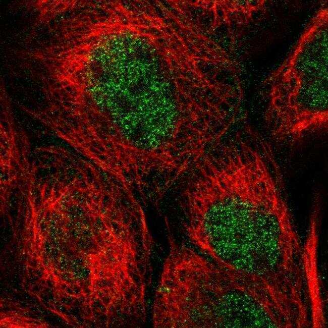
- Main image
- Experimental details
- Immunofluorescent staining of WIPI2 in human cell line A-431 shows positivity in nucleus but excluded from the nucleoli. Samples were probed using a WIPI2 Polyclonal Antibody (Product # PA5-54425).