Explore
 Validate
 Learn
Learn Western blot
 Immunocytochemistry
Antibody data
- Antibody Data
- Antigen structure
- References [3]
- Comments [0]
- Validations
- Western blot [8]
- Immunocytochemistry [1]
- Immunoprecipitation [1]
- Immunohistochemistry [1]
Submit
Validation data
Reference
Comment
Report error
- Product number
- GTX113266 - Provider product page

- Provider
- GeneTex
- Proper citation
- GeneTex Cat#GTX113266, RRID:AB_2037902
- Product name
- ROCK1 antibody [N1N2], N-term
- Antibody type
- Polyclonal
- Reactivity
- Human, Mouse, Rat
- Host
- Rabbit
Submitted references Cell surface flip-flop of phosphatidylserine is critical for PIEZO1-mediated myotube formation.
Androgen-regulated microRNA-135a decreases prostate cancer cell migration and invasion through downregulating ROCK1 and ROCK2.
Estrogen-related receptor α decreases RHOA stability to induce orientated cell migration.
Tsuchiya M, Hara Y, Okuda M, Itoh K, Nishioka R, Shiomi A, Nagao K, Mori M, Mori Y, Ikenouchi J, Suzuki R, Tanaka M, Ohwada T, Aoki J, Kanagawa M, Toda T, Nagata Y, Matsuda R, Takayama Y, Tominaga M, Umeda M
Nature communications 2018 May 24;9(1):2049
Nature communications 2018 May 24;9(1):2049
Androgen-regulated microRNA-135a decreases prostate cancer cell migration and invasion through downregulating ROCK1 and ROCK2.
Kroiss A, Vincent S, Decaussin-Petrucci M, Meugnier E, Viallet J, Ruffion A, Chalmel F, Samarut J, Allioli N
Oncogene 2015 May 28;34(22):2846-55
Oncogene 2015 May 28;34(22):2846-55
Estrogen-related receptor α decreases RHOA stability to induce orientated cell migration.
Sailland J, Tribollet V, Forcet C, Billon C, Barenton B, Carnesecchi J, Bachmann A, Gauthier KC, Yu S, Giguère V, Chan FL, Vanacker JM
Proceedings of the National Academy of Sciences of the United States of America 2014 Oct 21;111(42):15108-13
Proceedings of the National Academy of Sciences of the United States of America 2014 Oct 21;111(42):15108-13
No comments: Submit comment
Enhanced validation
Supportive validation
- Submitted by
- GeneTex (provider)
- Enhanced method
- Genetic validation
- Main image
- Experimental details
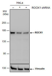
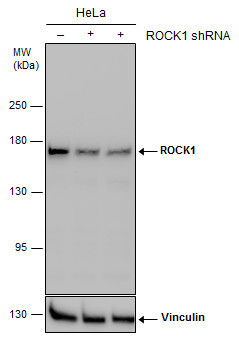
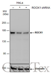
- Non-transfected (¡V) and transfected (+) HeLa whole cell extracts (30 ?g) were separated by 5% SDS-PAGE, and the membrane was blotted with ROCK1 antibody [N1N2], N-term (GTX113266) diluted at 1:1000. The HRP-conjugated anti-rabbit IgG antibody (GTX213110-01) was used to detect the primary antibody.
Supportive validation
- Submitted by
- GeneTex (provider)
- Main image
- Experimental details
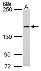
- ROCK1 antibody [N1N2], N-term detects ROCK1 protein by western blot analysis.A. 50 ?g rat brain lysate/extract5% SDS-PAGEROCK1 antibody [N1N2], N-term (GTX113266) dilution: 1:500 The HRP-conjugated anti-rabbit IgG antibody (GTX213110-01) was used to detect the primary antibody.

- Submitted by
- GeneTex (provider)
- Main image

- Experimental details
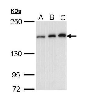
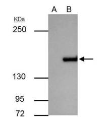
- Sample (30 ug of whole cell lysate) A: A431 (GTX27909) B: H1299 C: Molt-4 (GTX27912) 5% SDS PAGE GTX113266 diluted at 1:1000
- Validation comment
- WB
- Submitted by
- GeneTex (provider)
- Main image
- Experimental details
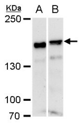
- ROCK1 antibody [N1N2], N-term detects ROCK1 protein by western blot analysis.A. 30 ?g NIH-3T3 whole cell extract B. 30 ?g C2Cl2 whole cell extract5% SDS-PAGEROCK1 antibody [N1N2], N-term (GTX113266) dilution: 1:1000 The HRP-conjugated anti-rabbit IgG antibody (GTX213110-01) was used to detect the primary antibody.

- Submitted by
- GeneTex (provider)
- Main image
- Experimental details
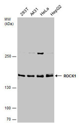
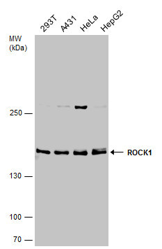
- Various whole cell extracts (30 ?g) were separated by 5% SDS-PAGE, and the membrane was blotted with ROCK1 antibody [N1N2], N-term (GTX113266) diluted at 1:1000. The HRP-conjugated anti-rabbit IgG antibody (GTX213110-01) was used to detect the primary antibody.
- Submitted by
- GeneTex (provider)
- Main image
- Experimental details
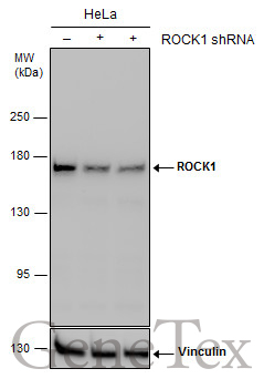
- Non-transfected (¡V) and transfected (+) HeLa whole cell extracts (30 ?g) were separated by 5% SDS-PAGE, and the membrane was blotted with ROCK1 antibody [N1N2], N-term (GTX113266) diluted at 1:1000. The HRP-conjugated anti-rabbit IgG antibody (GTX213110-01) was used to detect the primary antibody.
- Submitted by
- GeneTex (provider)
- Main image
- Experimental details
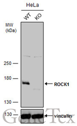
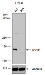
- Wild-type (WT) and ROCK1 knockout (KO) HeLa cell extracts (30 ?g) were separated by 5% SDS-PAGE, and the membrane was blotted with ROCK1 antibody [N1N2], N-term (GTX113266) diluted at 1:1000. The HRP-conjugated anti-rabbit IgG antibody (GTX213110-01) was used to detect the primary antibody.
- Submitted by
- GeneTex (provider)
- Main image
- Experimental details
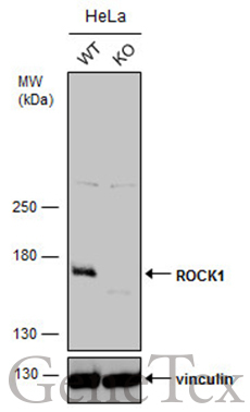
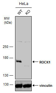
- Wild-type (WT) and ROCK1 knockout (KO) HeLa cell extracts (30 ?g) were separated by 5% SDS-PAGE, and the membrane was blotted with ROCK1 antibody [N1N2], N-term (GTX113266) diluted at 1:1000. The HRP-conjugated anti-rabbit IgG antibody (GTX213110-01) was used to detect the primary antibody.
Supportive validation
- Submitted by
- GeneTex (provider)
- Main image
- Experimental details
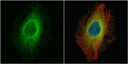
- ROCK1 antibody [N1N2], N-term detects ROCK1 protein at cytoplasm by immunofluorescent analysis.Sample: HeLa cells were fixed in 4% paraformaldehyde at RT for 15 min.Green: ROCK1 protein stained by ROCK1 antibody [N1N2], N-term (GTX113266) diluted at 1:500.Red: alpha Tubulin, a cytoskeleton marker, stained by alpha Tubulin antibody [GT114] (GTX628802) diluted at 1:500.Blue: Hoechst 33342 staining.
Supportive validation
- Submitted by
- GeneTex (provider)
- Main image
- Experimental details
- ROCK1 antibody immunoprecipitates ROCK1 protein in IP experiments. IP Sample: A431 whole cell lysate/extract A : Control with 3 £gg of pre-immune rabbit IgG B : Immunoprecipitation of ROCK1 by 3 £gg of ROCK1 antibody (GTX113266) 5% SDS-PAGE The immunoprecipitated ROCK1 protein was detected by ROCK1 antibody (GTX113266) diluted at 1 : 1000. EasyBlot anti-rabbit IgG (HRP) (GTX221666-01) was used as a secondary reagent.

Supportive validation
- Submitted by
- GeneTex (provider)
- Main image
- Experimental details
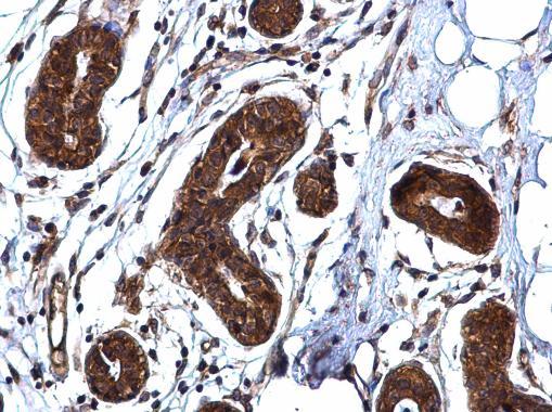
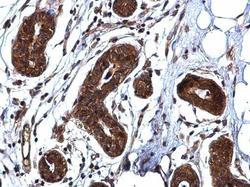
- ROCK1 antibody [N1N2], N-term detects ROCK1 protein at cytoplasm on human breast carcinoma by immunohistochemical analysis. Sample: Paraffin-embedded human breast carcinoma. ROCK1 antibody [N1N2], N-term (GTX113266) diluted at 1:500.