Explore
 Validate
 Learn
Learn Western blot
 Immunocytochemistry
Antibody data
- Antibody Data
- Antigen structure
- References [4]
- Comments [0]
- Validations
- Immunocytochemistry [2]
- Immunoprecipitation [1]
- Immunohistochemistry [1]
- Other assay [4]
Submit
Validation data
Reference
Comment
Report error
- Product number
- PA5-22262 - Provider product page

- Provider
- Invitrogen Antibodies
- Product name
- ROCK1 Polyclonal Antibody
- Antibody type
- Polyclonal
- Antigen
- Recombinant full-length protein
- Description
- Recommended positive controls: 293T, A431, HeLa, HepG2, NIH-3T3, C2Cl2, rat brain. Predicted reactivity: Human (98%), Mouse (98%), Rat (98%), Zebrafish (90%), Rabbit (98%), Chicken (96%), Rhesus Monkey (98%), Bovine (99%). Store product as a concentrated solution. Centrifuge briefly prior to opening the vial.
- Reactivity
- Human, Mouse, Rat
- Host
- Rabbit
- Isotype
- IgG
- Vial size
- 100 μL
- Concentration
- 0.35 mg/mL
- Storage
- Store at 4°C short term. For long term storage, store at -20°C, avoiding freeze/thaw cycles.
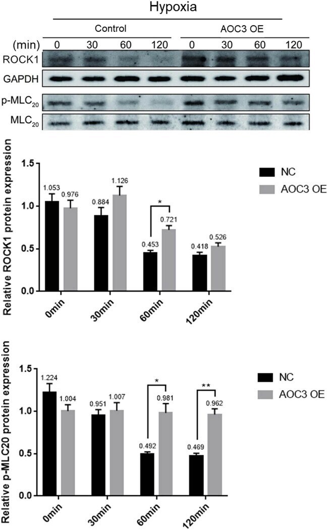
Submitted references Endothelial cell-derived SSAO can increase MLC(20) phosphorylation in VSMCs.
Circ_0057558 promotes nonalcoholic fatty liver disease by regulating ROCK1/AMPK signaling through targeting miR-206.
Periodic propagating waves coordinate RhoGTPase network dynamics at the leading and trailing edges during cell migration.
Rho-associated kinase 1 inhibition is synthetically lethal with von Hippel-Lindau deficiency in clear cell renal cell carcinoma.
Zhang Y, Zhang X, Cao Z, Huang Y, Zheng Y, Yang X
Open life sciences 2021;16(1):1141-1150
Open life sciences 2021;16(1):1141-1150
Circ_0057558 promotes nonalcoholic fatty liver disease by regulating ROCK1/AMPK signaling through targeting miR-206.
Chen X, Tan QQ, Tan XR, Li SJ, Zhang XX
Cell death & disease 2021 Aug 26;12(9):809
Cell death & disease 2021 Aug 26;12(9):809
Periodic propagating waves coordinate RhoGTPase network dynamics at the leading and trailing edges during cell migration.
Bolado-Carrancio A, Rukhlenko OS, Nikonova E, Tsyganov MA, Wheeler A, Garcia-Munoz A, Kolch W, von Kriegsheim A, Kholodenko BN
eLife 2020 Jul 24;9
eLife 2020 Jul 24;9
Rho-associated kinase 1 inhibition is synthetically lethal with von Hippel-Lindau deficiency in clear cell renal cell carcinoma.
Thompson JM, Nguyen QH, Singh M, Pavesic MW, Nesterenko I, Nelson LJ, Liao AC, Razorenova OV
Oncogene 2017 Feb 23;36(8):1080-1089
Oncogene 2017 Feb 23;36(8):1080-1089
No comments: Submit comment
Supportive validation
- Submitted by
- Invitrogen Antibodies (provider)
- Main image
- Experimental details
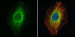
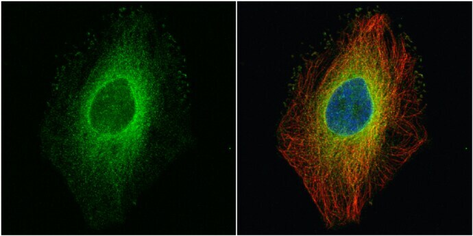
- Immunocytochemistry-Immunofluorescence analysis of ROCK1 was performed in HeLa cells fixed in 4% paraformaldehyde at RT for 15 min. Green: ROCK1 Polyclonal Antibody (Product # PA5-22262) diluted at 1:500. Red: alpha Tubulin, a cytoskeleton marker. Blue: Hoechst 33342 staining.
- Submitted by
- Invitrogen Antibodies (provider)
- Main image

- Experimental details
- Immunocytochemistry-Immunofluorescence analysis of ROCK1 was performed in HeLa cells fixed in 4% paraformaldehyde at RT for 15 min. Green: ROCK1 Polyclonal Antibody (Product # PA5-22262) diluted at 1:500. Red: alpha Tubulin, a cytoskeleton marker. Blue: Hoechst 33342 staining.

Supportive validation
- Submitted by
- Invitrogen Antibodies (provider)
- Main image

- Experimental details
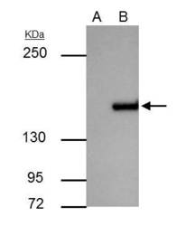
- ROCK1 antibody immunoprecipitates ROCK1 protein in IP experiments. IP Sample: A431 whole cell lysate/extract A : Control with 3 µg of pre-immune rabbit IgG B : Immunoprecipitation of ROCK1 by 3 µg of ROCK1 antibody (Product # PA5-22262) 5% SDS-PAGE The immunoprecipitated ROCK1 protein was detected by ROCK1 antibody (Product # PA5-22262) diluted at 1 : 500. Anti-rabbit IgG (HRP) was used as a secondary reagent.

Supportive validation
- Submitted by
- Invitrogen Antibodies (provider)
- Main image
- Experimental details
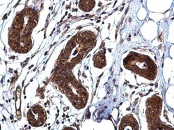
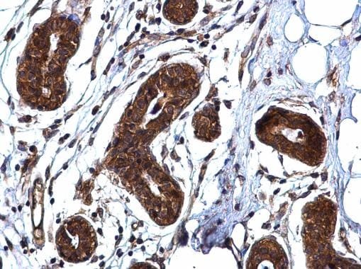
- ROCK1 Polyclonal Antibody detects ROCK1 protein at cytoplasm on human breast carcinoma by immunohistochemical analysis. Sample: Paraffin-embedded human breast carcinoma. ROCK1 Polyclonal Antibody (Product # PA5-22262) diluted at 1:500. Antigen Retrieval: EDTA based buffer, pH 8.0, 15 min.
Supportive validation
- Submitted by
- Invitrogen Antibodies (provider)
- Main image
- Experimental details
- ROCK1 antibody immunoprecipitates ROCK1 protein in IP experiments. IP Sample: A431 whole cell lysate/extract A : Control with 3 µg of pre-immune rabbit IgG B : Immunoprecipitation of ROCK1 by 3 µg of ROCK1 antibody (Product # PA5-22262) 5% SDS-PAGE The immunoprecipitated ROCK1 protein was detected by ROCK1 antibody (Product # PA5-22262) diluted at 1 : 500. Anti-rabbit IgG (HRP) was used as a secondary reagent.

- Submitted by
- Invitrogen Antibodies (provider)
- Main image
- Experimental details
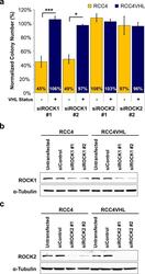
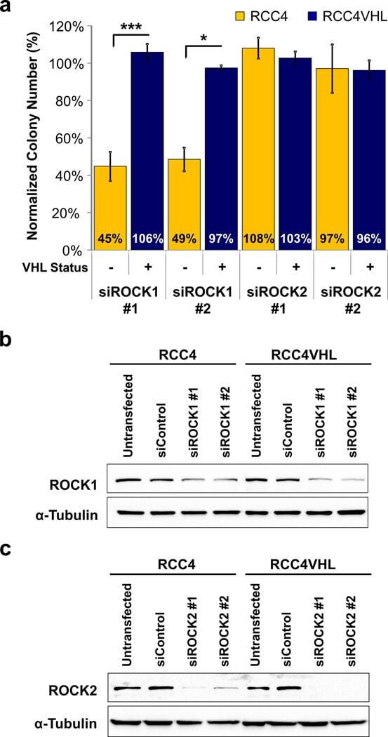
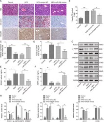
- Figure 2 Synthetic lethality of Y-27632 with VHL loss is mimicked by siRNA downregulation of ROCK1, not ROCK2 RCC4+-VHL matched cell lines were transfected with siRNAs targeting ROCK1, ROCK2, or non-targeting siRNA control (siControl). Twenty-four hours after transfection cells were plated for a clonogenic assay. Each transfection was done in triplicate, followed by clonogenic assays conducted in triplicate, and the experiments were repeated at least two times. ( a ) Transfection with siROCK1, but not siROCK2, resulted in significant reduction in RCC4 colony numbers in comparison to RCC4VHL. Thus, ROCK1 downregulation mimics the effect of Y-27632 treatment on viability of RCC4 cells, making it a likely target for Y-27632 causing synthetic lethality effect. Statistical analysis was performed using a paired t-test comparing numbers of colonies in each siROCK group to siControl. SEMs are shown. (b-c) The degree of each target knockdown by its specific siRNA (as indicated) was assessed by Western blot.
- Submitted by
- Invitrogen Antibodies (provider)
- Main image
- Experimental details
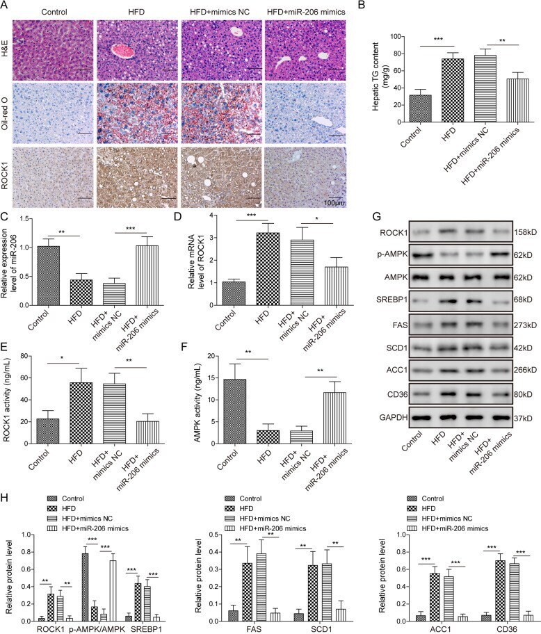
- Fig. 2 miR-206 inhibited lipogenesis and TG secretion in vivo. A Representative images of H&E staining (upper), oil-red o staining (middle), and IHC staining (bottom) in liver tissues from HFD-fed mice or control mice with injection of miR-206 mimics or NC. B Secreted TG levels in liver tissues from HFD-fed mice or control mice with injection of miR-206 mimics or NC. C miR-206 levels in liver tissues from HFD-fed mice or control mice with injection of miR-206 mimics or NC. D ROCK1 mRNA levels in liver tissues from HFD-fed mice or control mice with injection of miR-206 mimics or NC. E , F ROCK1/AMPK activities in liver tissues from HFD-fed mice or control mice with injection of miR-206 mimics or NC. G Protein levels of ROCK1, p-AMPK, and lipogenesis-related proteins in liver tissues from HFD-fed mice or control mice with injection of miR-206 mimics or NC. H Quantifications of G . * p < 0.05, ** p < 0.01, and *** p < 0.001.
- Submitted by
- Invitrogen Antibodies (provider)
- Main image
- Experimental details
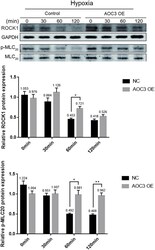
- Figure 4 Overexpression of AOC3 increases the ROCK1 expression and phosphorylation of MLC 20 in RIMSCs under hypoxia. ROCK1 and p-MLC 20 protein expression in RIMSCs were determined by western blotting. GAPDH and MLC 20 were used as internal controls, respectively (* P < 0.05, ** P < 0.01, and *** P < 0.001).