Explore
 Validate
 Learn
LearnPA1-26351
antibody from Invitrogen Antibodies
Targeting: AURKA
AIK, ARK1, AurA, BTAK, PPP1R47, STK15, STK6, STK7
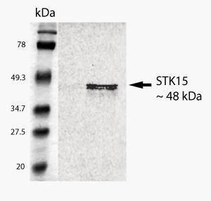
 Western blot
 Immunocytochemistry
 Immunohistochemistry
Antibody data
- Antibody Data
- Antigen structure
- References [1]
- Comments [0]
- Validations
- Immunocytochemistry [2]
- Immunoprecipitation [2]
- Other assay [1]
Submit
Validation data
Reference
Comment
Report error
- Product number
- PA1-26351 - Provider product page

- Provider
- Invitrogen Antibodies
- Product name
- Aurora A Polyclonal Antibody
- Antibody type
- Polyclonal
- Antigen
- Synthetic peptide
- Description
- PA1-26351 detects Aurora A from human samples.
- Reactivity
- Human
- Host
- Rabbit
- Isotype
- IgG
- Vial size
- 50 μg
- Concentration
- 1 mg/mL
- Storage
- 4°C
Submitted references A kinase-independent function for AURORA-A in replisome assembly during DNA replication initiation.
Guarino Almeida E, Renaudin X, Venkitaraman AR
Nucleic acids research 2020 Aug 20;48(14):7844-7855
Nucleic acids research 2020 Aug 20;48(14):7844-7855
No comments: Submit comment
Supportive validation
- Submitted by
- Invitrogen Antibodies (provider)
- Main image
- Experimental details
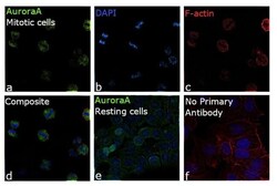
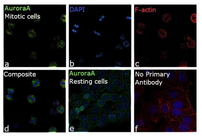
- Immunofluorescence analysis of Aurora A was performed using 70% confluent log phase MCF7 cells. The cells were fixed with 4% paraformaldehyde for 10 minutes, permeabilized with 0.1% Triton™ X-100 for 15 minutes, and blocked with 2% BSA for 1 hour at room temperature. The cells were labeled with Aurora A Polyclonal Antibody (Product # PA1-26351) at 5 µg/mL concentration in 0.1% BSA, incubated at 4 degree celsius overnight (Panel a: green). Mitotic chromosomes (Panel b: blue) were stained with ProLong™ Diamond Antifade Mountant with DAPI (Product # P36962). F-actin (Panel c: red) was stained with Rhodamine Phalloidin (Product # R415, 1:300). Panel d represents the merged image showing mitotic cells with AuroraA expression concentrated at the spindle poles. Panel e represents the resting cells showing cytoplasmic expression of AuroraA . Panel f represents control cells with no primary antibody to assess background. The images were captured at 60X magnification.
- Submitted by
- Invitrogen Antibodies (provider)
- Main image

- Experimental details
- Immunofluorescence analysis of Aurora A was performed using 70% confluent log phase MCF7 cells. The cells were fixed with 4% paraformaldehyde for 10 minutes, permeabilized with 0.1% Triton™ X-100 for 15 minutes, and blocked with 2% BSA for 1 hour at room temperature. The cells were labeled with Aurora A Polyclonal Antibody (Product # PA1-26351) at 5 µg/mL concentration in 0.1% BSA, incubated at 4 degree celsius overnight (Panel a: green). Mitotic chromosomes (Panel b: blue) were stained with ProLong™ Diamond Antifade Mountant with DAPI (Product # P36962). F-actin (Panel c: red) was stained with Rhodamine Phalloidin (Product # R415, 1:300). Panel d represents the merged image showing mitotic cells with AuroraA expression concentrated at the spindle poles. Panel e represents the resting cells showing cytoplasmic expression of AuroraA . Panel f represents control cells with no primary antibody to assess background. The images were captured at 60X magnification.

Supportive validation
- Submitted by
- Invitrogen Antibodies (provider)
- Main image
- Experimental details
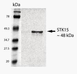
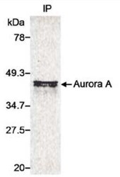
- Immunoprecipitation of S35 labeled recombinant Aurora A (5.0 mg) using Aurora A polyclonal antibody (Product # PA1-26351) followed by gel electrophoresis.
- Submitted by
- Invitrogen Antibodies (provider)
- Main image
- Experimental details
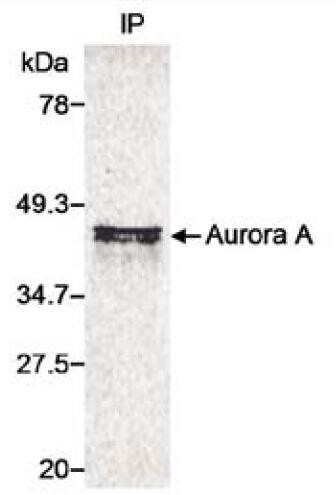
- IP analysis of 35S-Met-labeled whole cell lysate transfected with a human Aurora A expression construct using (Product # PA1-26351) Aurora A Polyclonal Antibody.IP reaction : 2-10 µg/mg lysate.

Supportive validation
- Submitted by
- Invitrogen Antibodies (provider)
- Main image

- Experimental details
- IP analysis of 35S-Met-labeled whole cell lysate transfected with a human Aurora A expression construct using (Product # PA1-26351) Aurora A Polyclonal Antibody.IP reaction : 2-10 µg/mg lysate.