Explore
 Validate
 Learn
LearnPA5-34700
antibody from Invitrogen Antibodies
Targeting: AURKA
AIK, ARK1, AurA, BTAK, PPP1R47, STK15, STK6, STK7
 Western blot
Antibody data
- Antibody Data
- Antigen structure
- References [0]
- Comments [0]
- Validations
- Western blot [6]
- Immunocytochemistry [4]
- Immunohistochemistry [1]
Submit
Validation data
Reference
Comment
Report error
- Product number
- PA5-34700 - Provider product page

- Provider
- Invitrogen Antibodies
- Product name
- Aurora A Polyclonal Antibody
- Antibody type
- Polyclonal
- Antigen
- Synthetic peptide
- Description
- Recommended positive controls: Aurora A-transfected 293T. Predicted reactivity: Chimpanzee (100%). Store product as a concentrated solution. Centrifuge briefly prior to opening the vial.
- Reactivity
- Human
- Host
- Rabbit
- Isotype
- IgG
- Vial size
- 100 µL
- Concentration
- 0.87 mg/mL
- Storage
- Store at 4°C short term. For long term storage, store at -20°C, avoiding freeze/thaw cycles.
No comments: Submit comment
Supportive validation
- Submitted by
- Invitrogen Antibodies (provider)
- Main image

- Experimental details
- Western blot analysis of Aurora A using A) 30 µg 293T whole cell lysate (B) 30 µg A431 whole cell lysate (C) 30 µg HeLa whole cell lysate (D) 30 µg HepG2 whole cell lysate and E) 30 µg A375 whole cell lysate. Samples were loaded onto a 10% SDS-PAGE gel and probed with an Aurora A polyclonal antibody (Product # PA5-34700) at a dilution of 1:1000.

- Submitted by
- Invitrogen Antibodies (provider)
- Main image

- Experimental details
- Western blot analysis of Aurora A using A) 30 µg Neuro2A whole cell lysate and B) 30 µg GL261 whole cell lysate. Samples were loaded onto a 10% SDS-PAGE gel and probed with an Aurora A polyclonal antibody (Product # PA5-34700) at a dilution of 1:1000.

- Submitted by
- Invitrogen Antibodies (provider)
- Main image

- Experimental details
- Western blot analysis of Aurora A using 30 µg PC-12 whole cell lysate. Samples were loaded onto a 10% SDS-PAGE gel and probed with an Aurora A polyclonal antibody (Product # PA5-34700) at a dilution of 1:1000.

- Submitted by
- Invitrogen Antibodies (provider)
- Main image

- Experimental details
- Western Blot using Aurora A Polyclonal Antibody (Product # PA5-34700). Non-transfected (–) and transfected (+) 293T whole cell extracts (30 µg) were separated by 10% SDS-PAGE, and the membrane was blotted with Aurora A Polyclonal Antibody (Product # PA5-34700) diluted at 1:1,000. The HRP-conjugated anti-rabbit IgG antibody was used to detect the primary antibody.

- Submitted by
- Invitrogen Antibodies (provider)
- Main image

- Experimental details
- Western Blot analysis of Aurora A was performed by separating 30 µg of non-transfected (–) and transfected (+) 293T whole cell extracts by 10% SDS-PAGE. Proteins were transferred to a membrane and probed with a Aurora A Polyclonal Antibody (Product # PA5-34700) at a dilution of 1:1000. The HRP-conjugated anti-rabbit IgG antibody was used to detect the primary antibody.

- Submitted by
- Invitrogen Antibodies (provider)
- Main image

- Experimental details
- Western Blot using Aurora A Polyclonal Antibody (Product # PA5-34700). Non-transfected (–) and transfected (+) 293T whole cell extracts (30 µg) were separated by 10% SDS-PAGE, and the membrane was blotted with Aurora A Polyclonal Antibody (Product # PA5-34700) diluted at 1:1,000. The HRP-conjugated anti-rabbit IgG antibody was used to detect the primary antibody.

Supportive validation
- Submitted by
- Invitrogen Antibodies (provider)
- Main image
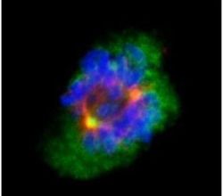
- Experimental details
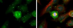
- Immunofluorescent analysis of Aurora an In methanol-fixed 293T cells using an Aurora A polyclonal antibody (Product # PA5-34700) (Green) at a 1:500 dilution. Alpha-tubulin filaments were labeled with Product # PA5-29281 (Red) at a 1:2500.

- Submitted by
- Invitrogen Antibodies (provider)
- Main image
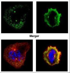
- Experimental details
- Immunofluorescent analysis of Aurora an In paraformaldehyde-fixed U2OS cells using an Aurora A polyclonal antibody (Product # PA5-34700) (Green) at a 1:500 dilution. Alpha-tubulin filaments were labeled with Product # PA5-29281 (Red) at a 1:500.

- Submitted by
- Invitrogen Antibodies (provider)
- Main image
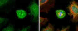
- Experimental details
- Immunocytochemistry-Immunofluorescence analysis of Aurora A was performed in HeLa cells fixed in ice cold MeOH for 5 min. Green: Aurora A Polyclonal Antibody (Product # PA5 34700) diluted at 1:500. Red: alpha Tubulin, a cytoskeleton marker. Blue: Hoechst 33342 staining.

- Submitted by
- Invitrogen Antibodies (provider)
- Main image
- Experimental details
- Immunocytochemistry-Immunofluorescence analysis of Aurora A was performed in HeLa cells fixed in ice cold MeOH for 5 min. Green: Aurora A Polyclonal Antibody (Product # PA5 34700) diluted at 1:500. Red: alpha Tubulin, a cytoskeleton marker. Blue: Hoechst 33342 staining.

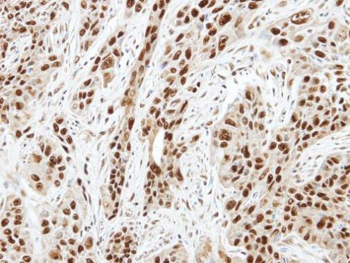
Supportive validation
- Submitted by
- Invitrogen Antibodies (provider)
- Main image
- Experimental details
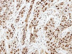
- Immunohistochemical analysis of paraffin-embedded TW2.6 xenograft, using Aurora A (Product # PA5-34700) antibody at 1:100 dilution. Antigen Retrieval: EDTA based buffer, pH 8.0, 15 min.