 Explore
 Validate
 Learn
LearnSTJ99106
antibody from St John's Laboratory
Targeting: AURKA
AIK, ARK1, AurA, BTAK, PPP1R47, STK15, STK6, STK7
 Western blot
Antibody data
- Antibody Data
- Antigen structure
- References [0]
- Comments [0]
- Validations
- Western blot [1]
- Immunocytochemistry [1]
- Other assay [1]
Submit
Validation data
Reference
Comment
Report error
- Product number
- STJ99106 - Provider product page

- Provider
- St John's Laboratory
- Product name
- Anti-AURKA antibody [2E3-C10-C11-G6] (STJ99106)
- Antibody type
- Monoclonal
- Description
- Mouse monoclonal antibody anti-Aurora Kinase A is suitable for use in Western Blot, Immunofluorescence and Immunocytochemistry research applications.
- Reactivity
- Human, Simian
- Host
- Mouse
- Conjugate
- Unconjugated
- Antigen sequence
NA- Epitope
- NA
- Isotype
- IgG
- Antibody clone number
- NA
- Vial size
- NA
- Concentration
- NA
- Storage
- Store at-20°C for up to 1 year from the date of receipt, and avoid repeat freeze-thaw cycles.
- Handling
- NA
No comments: Submit comment
Supportive validation
- Submitted by
- St John's Laboratory (provider)
- Main image
- Experimental details
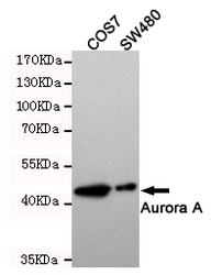
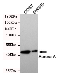
- Western blot detection of Aurora Kinase A in SW480 and COS7 cell lysates and using Aurora Kinase A mouse mAb (1:500 diluted).Predicted band size: 46KDa.Observed band size:46KDa.
- Sample type
- NA
- Validation comment
- NA
- Primary Ab dilution
- NA
- Other comments
- NA
- Secondary Ab
- NA
- Secondary Ab dilution
- NA
- Protocol
- NA
Supportive validation
- Submitted by
- St John's Laboratory (provider)
- Main image
- Experimental details
- Immunocytochemistry staining of HeLa cells fixed with-20℃ Methanol and using Aurora Kinase A mouse mAb (dilution 1:100).
- Sample type
- NA
- Validation comment
- NA
- Primary Ab dilution
- NA
- Other comments
- NA
- Secondary Ab
- NA
- Secondary Ab dilution
- NA
- Protocol
- NA
Supportive validation
- Submitted by
- St John's Laboratory (provider)
- Main image
- Experimental details
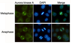
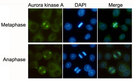
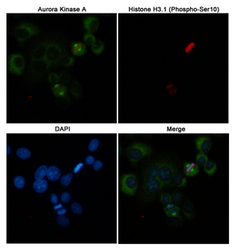
- Immunofluorescent analysis of Hela cells labeled with Aurora Kinase A (200525, dilution 1:50) mouse mAb (green) and Histone H3.1 (Phospho-Ser10) (310045, dilution 1:100) Rabbit pAb (red). DAPI was used to stain nucleus (blue).
- Sample type
- NA
- Validation comment
- NA
- Primary Ab dilution
- NA
- Other comments
- NA
- Secondary Ab
- NA
- Secondary Ab dilution
- NA
- Protocol
- NA
