 Explore
 Validate
 Learn
Learn Western blot
 Immunocytochemistry
Antibody data
- Antibody Data
- Antigen structure
- References [1]
- Comments [0]
- Validations
- Western blot [1]
Submit
Validation data
Reference
Comment
Report error
- Product number
- 603302 - Provider product page

- Provider
- BioLegend
- Proper citation
- BioLegend Cat#603302, RRID:AB_315623
- Product name
- Purified anti-Aurora A (Aurora 2)
- Antibody type
- Polyclonal
- Antigen
- Recombinant (partial), N-terminal
- Description
- Aurora A (also known as Aurora 2) is a serine/threonine kinase with a molecular weight of approximately 46 kD. This kinase is highly expressed in the thymus and some tumors and is also expressed in other tissues including the lung, testis, colon, placenta, and fetal liver. Aurora A localizes in the midzone or central spindle in late anaphase and is concentrated in the midbody in telophase and during cytokinesis. This kinase is believed to act in cell cycle regulation during anaphase and/or telophase at centrosome/spindle pole during chromosome segregation. Aurora A has been shown to regulate cleavage of polar spindle microtubules at the onset of cytokinesis during mitosis. Defects in Aurora A cause numerous centrosome aberrations including aneuploidy (genetic variant with amino acid substitution F31I). Aurora A expression is cell cycle regulated, low in G1/S, and accumulating in G2/M. Expression is upregulated in cancer cells during M phase. Phosphorylation by PKA has been shown to regulate function. Aurora A phosphorylation has been reported on Thr 288. This kinase associates with the centrosome and mitotic spindles, NM23-H1, protein phosphatase type I, and co-localizes with γ-tubulin. The Phe 31 variant has been shown to interact with the E2 ubiquitin-conjugating enzyme, UBE2N. The Poly6033 antibody recognizes the N-terminal region of Aurora A. The Poly6033 antibody has been shown to be useful for Western blotting of human and mouse Aurora A protein and for immunoprecipitation of human Aurora A (mouse not tested).
- Reactivity
- Human, Mouse
- Host
- Rabbit
- Conjugate
- Unconjugated
- Vial size
- 200 µl
- Storage
- Upon receipt, store frozen at -20° C.
Submitted references Specialized roles of the two mitotic cyclins in somatic cells: cyclin A as an activator of M phase-promoting factor.
Fung TK, Ma HT, Poon RY
Molecular biology of the cell 2007 May;18(5):1861-73
Molecular biology of the cell 2007 May;18(5):1861-73
No comments: Submit comment
Supportive validation
- Submitted by
- BioLegend (provider)
- Main image
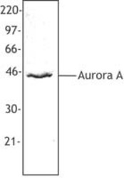
- Experimental details
- Hela cell nuclear extract was resolved by electrophoresis, transferred to nitrocellulose, and probed with rabbit anti-Aurora A antibody. Proteins were visualized using a donkey anti-rabbit secondary conjugated to HRP and a chemiluminescence detection system.
- Conjugate
- Unconjugated
