Explore
 Validate
 Learn
LearnNB100-267
antibody from Novus Biologicals
Targeting: AURKA
AIK, ARK1, AurA, BTAK, PPP1R47, STK15, STK6, STK7
 Western blot
Antibody data
- Antibody Data
- Antigen structure
- References [5]
- Comments [0]
- Validations
- Western blot [4]
- Immunocytochemistry [1]
- Immunoprecipitation [1]
Submit
Validation data
Reference
Comment
Report error
- Product number
- NB100-267 - Provider product page

- Provider
- Novus Biologicals
- Proper citation
- Novus Cat#NB100-267, RRID:AB_10002481
- Product name
- Rabbit Polyclonal Aurora A Antibody
- Antibody type
- Polyclonal
- Description
- Immunogen affinity purified. This is specific for human STK15, Serine/Threonine Kinase 15, Aurora 2, BRAK, ARK1, Aurora-Related Kinase 1
- Reactivity
- Human
- Host
- Rabbit
- Isotype
- IgG
- Vial size
- 100 ul
- Concentration
- 1.0 mg/ml
- Storage
- Store at 4C. Do not freeze.
Submitted references The nuclear scaffold protein SAF-A is required for kinetochore-microtubule attachment and contributes to the targeting of Aurora-A to mitotic spindles.
RASSF1A interacts with and activates the mitotic kinase Aurora-A.
A role for IkappaB kinase 2 in bipolar spindle assembly.
Overexpression of the centrosomal protein Aurora-A kinase is associated with poor prognosis in epithelial ovarian cancer patients.
Aurora a and B overexpression and centrosome amplification in early estrogen-induced tumor foci in the Syrian hamster kidney: implications for chromosomal instability, aneuploidy, and neoplasia.
Ma N, Matsunaga S, Morimoto A, Sakashita G, Urano T, Uchiyama S, Fukui K
Journal of cell science 2011 Feb 1;124(Pt 3):394-404
Journal of cell science 2011 Feb 1;124(Pt 3):394-404
RASSF1A interacts with and activates the mitotic kinase Aurora-A.
Liu L, Guo C, Dammann R, Tommasi S, Pfeifer GP
Oncogene 2008 Oct 16;27(47):6175-86
Oncogene 2008 Oct 16;27(47):6175-86
A role for IkappaB kinase 2 in bipolar spindle assembly.
Irelan JT, Murphy TJ, DeJesus PD, Teo H, Xu D, Gomez-Ferreria MA, Zhou Y, Miraglia LJ, Rines DR, Verma IM, Sharp DJ, Tergaonkar V, Chanda SK
Proceedings of the National Academy of Sciences of the United States of America 2007 Oct 23;104(43):16940-5
Proceedings of the National Academy of Sciences of the United States of America 2007 Oct 23;104(43):16940-5
Overexpression of the centrosomal protein Aurora-A kinase is associated with poor prognosis in epithelial ovarian cancer patients.
Landen CN Jr, Lin YG, Immaneni A, Deavers MT, Merritt WM, Spannuth WA, Bodurka DC, Gershenson DM, Brinkley WR, Sood AK
Clinical cancer research : an official journal of the American Association for Cancer Research 2007 Jul 15;13(14):4098-104
Clinical cancer research : an official journal of the American Association for Cancer Research 2007 Jul 15;13(14):4098-104
Aurora a and B overexpression and centrosome amplification in early estrogen-induced tumor foci in the Syrian hamster kidney: implications for chromosomal instability, aneuploidy, and neoplasia.
Hontz AE, Li SA, Lingle WL, Negron V, Bruzek A, Salisbury JL, Li JJ
Cancer research 2007 Apr 1;67(7):2957-63
Cancer research 2007 Apr 1;67(7):2957-63
No comments: Submit comment
Supportive validation
- Submitted by
- Novus Biologicals (provider)
- Main image

- Experimental details
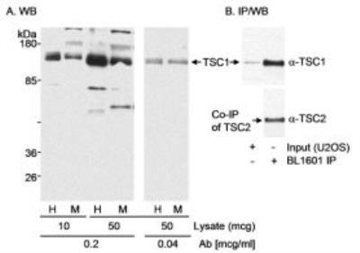
- Western Blot: Aurora A Antibody [NB100-267] - Detection of Human and Mouse TSC1.
- Submitted by
- Novus Biologicals (provider)
- Main image

- Experimental details
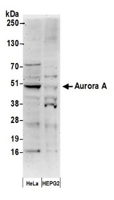
- Western Blot: Aurora A Antibody [NB100-267] - Samples: Whole cell lysate (50 ug) from HeLa and HEPG2cells prepared using NETN lysis buffer. Antibody: Affinity purified rabbit anti-Aurora A antibody used for WB at 0.2 ug/ml. Detection: Chemiluminescence with an exposure time of 3 minutes.
- Submitted by
- Novus Biologicals (provider)
- Main image
- Experimental details
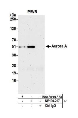
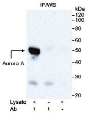
- Western Blot: Aurora A Antibody [NB100-267] - Detection of Human Aurora A (STK15) on HeLa whole cell lysate using NB100-267. Immunoprecipitated Aurora A was detected using a monoclonal antibody to Aurora A from another source.
- Submitted by
- Novus Biologicals (provider)
- Main image
- Experimental details
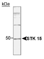
- Western Blot: Aurora A Antibody [NB100-267] - Western blot and Immunoprecipitation detection of human 5TK15, using NB100-267. Samples: Whole cell lysate from 5KBr3 (A) or AsPC-1 (B) cells.

Supportive validation
- Submitted by
- Novus Biologicals (provider)
- Main image

- Experimental details
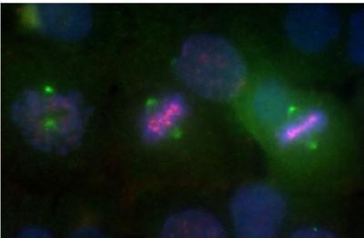
- Immunocytochemistry/Immunofluorescence: Aurora A Antibody [NB100-267] - Localization of Aurora A to Centrosomes asynchronous HeLa cells. NB 100-267 is diluted 1:1000. Aurora A(green) was visualized with FITC. DAPI (blue), Aurora B (pink).
Supportive validation
- Submitted by
- Novus Biologicals (provider)
- Main image
- Experimental details
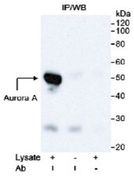
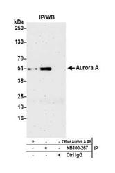
- Immunoprecipitation: Aurora A Antibody [NB100-267] - Detection of human Aurora A by western blot of immunoprecipitates. Samples: Whole cell lysate (1.0 mg per IP reaction; 20% of IP loaded) from HeLa cells prepared using NETN lysis buffer. Antibodies: Affinity purified rabbit anti-Aurora A antibody NB100-267 used for IP at 3 ug per reaction. Aurora A was also immunoprecipitated by another rabbit anti-Aurora A antibody. For blotting immunoprecipitated Aurora A, NB100-267 was used at 1 ug/ml. Detection: Chemiluminescence with an exposure time of 3 minutes.