Explore
 Validate
 Learn
Learn Western blot
 Immunohistochemistry
Antibody data
- Antibody Data
- Antigen structure
- References [1]
- Comments [0]
- Validations
- Immunohistochemistry [1]
- Other assay [1]
Submit
Validation data
Reference
Comment
Report error
- Product number
- MA5-27845 - Provider product page

- Provider
- Invitrogen Antibodies
- Product name
- CXCL16 Monoclonal Antibody (GT516)
- Antibody type
- Monoclonal
- Antigen
- Recombinant full-length protein
- Description
- Positive Control: HeLa, HeLa membrane extract Store product as a concentrated solution. Centrifuge briefly prior to opening the vial.
- Reactivity
- Human, Mouse, Rat
- Host
- Mouse
- Isotype
- IgG
- Antibody clone number
- GT516
- Vial size
- 100 μL
- Concentration
- 1 mg/mL
- Storage
- Store at 4°C short term. For long term storage, store at -20°C, avoiding freeze/thaw cycles.
Submitted references Dendritic cells maintain anti-tumor immunity by positioning CD8 skin-resident memory T cells.
Vella JL, Molodtsov A, Angeles CV, Branchini BR, Turk MJ, Huang YH
Life science alliance 2021 Oct;4(10)
Life science alliance 2021 Oct;4(10)
No comments: Submit comment
Supportive validation
- Submitted by
- Invitrogen Antibodies (provider)
- Main image
- Experimental details
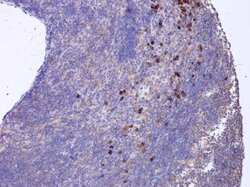
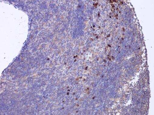
- CXCL16 Monoclonal Antibody (GT516) detects CXCL16 protein at cytoplasm in rat lymph node by immunohistochemical analysis. Sample: Paraffin-embedded rat lymph node. CXCL16 Monoclonal Antibody (GT516) (Product # MA5-27845) diluted at 1:200. Antigen Retrieval: Citrate buffer, pH 6.0, 15 min.
Supportive validation
- Submitted by
- Invitrogen Antibodies (provider)
- Main image
- Experimental details
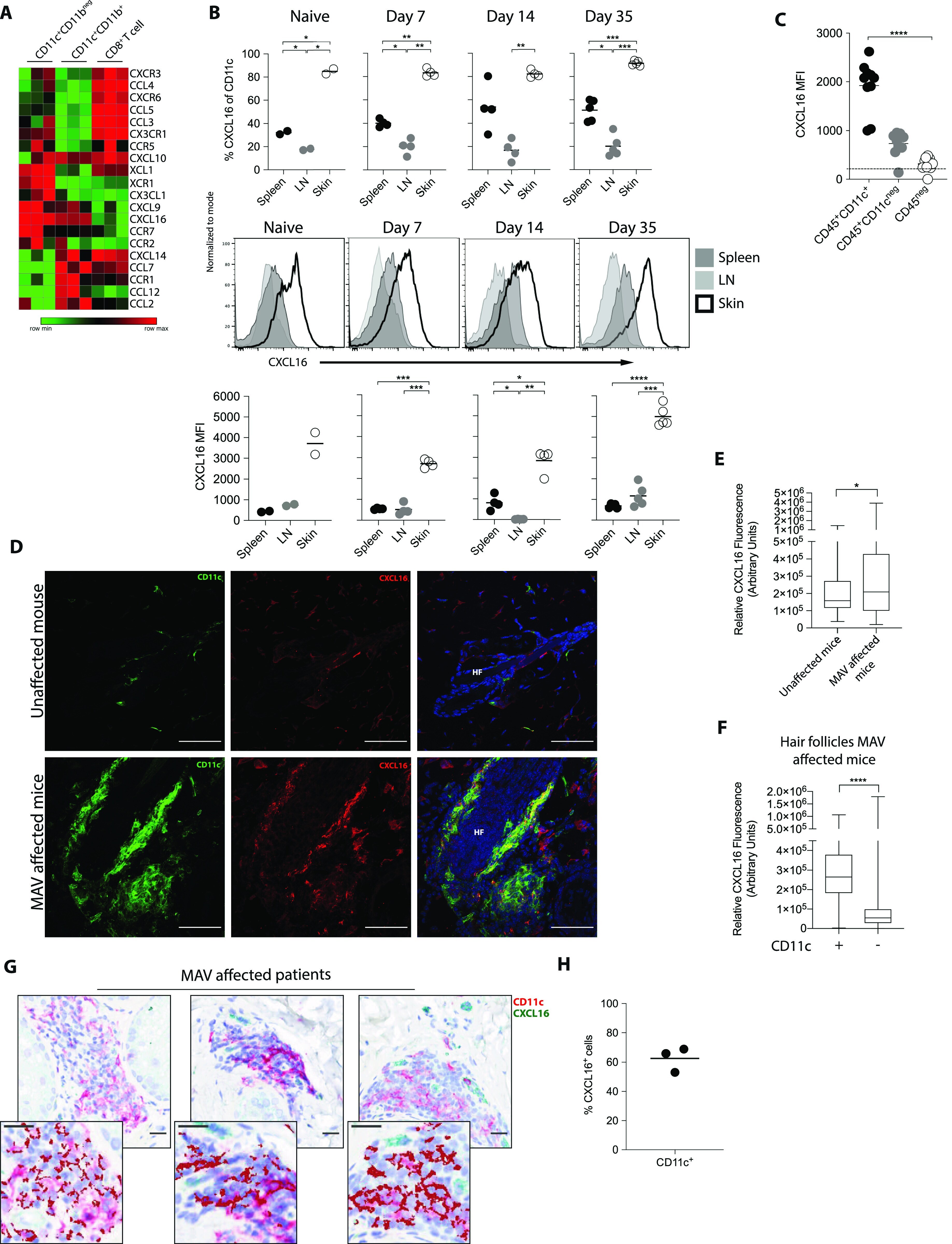
- Figure 3. CD11c + cells express CXCL16 in melanoma-associated vitiligo (MAV)-affected skin. (A) Heat map showing gene expression of chemokines and receptors in CD11c + CD11b neg , CD11c + CD11b + , and CD8 + T cells sorted from skin of MAV-affected mice. Data presented as log2-normalized expression. (B) Surface expression of CXCL16 on CD11c + cells isolated from skin of naive (n = 3) or MAV-affected (n = 5) mice, skin-draining LNs, or spleen 7, 14, and 35 d after surgery. Representative of three independent experiments. (C) Surface expression of CXCL16 on CD45 + CD11c + , CD45 + CD11c neg , and CD45 neg cells isolated from skin of MAV-affected mice, 35 d after surgery. Representative of two independent experiments with skin collected between 35- and 60-d post-surgery. Dotted horizontal line indicates background CXCL16 expression in unstained cell sample. (D) Expression of CXCL16 and CD11c in unaffected and MAV-affected mice; CD11c (green), CXCL16 (red), and nuclei (blue). Scale bar, 50 mum. (E) CXCL16 expression in IF images from skin of unaffected and MAV-affected mice; n = 5-8 mice. (F) CXCL16 expression in hair follicles with or without CD11c + cell clusters in MAV-affected skin; n = 8 mice. (G) CD11c + and CXCL16 + cells in skin from MAV-affected patients. Stains identify CXCL16 (green) and CD11c (red); dark red identifies colocalization of CD11c and CXCL16. Scale bar, 25 mum. (H) Percent of CD11c + cells expressing CXCL16 in MAV-affected patient skin. (B, C, H) Symbols r