Explore
 Validate
 Learn
Learn Immunoprecipitation
Antibody data
- Antibody Data
- Antigen structure
- References [0]
- Comments [0]
- Validations
- Immunoprecipitation [1]
- Immunohistochemistry [4]
- Other assay [4]
Submit
Validation data
Reference
Comment
Report error
- Product number
- PA5-51736 - Provider product page

- Provider
- Invitrogen Antibodies
- Product name
- BAZ1A Polyclonal Antibody
- Antibody type
- Polyclonal
- Antigen
- Recombinant protein fragment
- Description
- Immunogen sequence: RGHRESALKE TLLQEKSRIC AQLARFSEEK FHFSDKPQPD SKPTYSRGRS SNAYDPSQMC AEKQLELRLR DFLLDIEDRI YQGTLGAIKV TDRHIWRSAL ESGRYELLSE ENKENGIIKT VNEDVEEMEI DEQTK Highest antigen sequence identity to the following orthologs: Mouse - 85%, Rat - 83%.
- Reactivity
- Human
- Host
- Rabbit
- Isotype
- IgG
- Vial size
- 100 μL
- Concentration
- 0.1 mg/mL
- Storage
- Store at 4°C short term. For long term storage, store at -20°C, avoiding freeze/thaw cycles.
No comments: Submit comment
Supportive validation
- Submitted by
- Invitrogen Antibodies (provider)
- Main image

- Experimental details
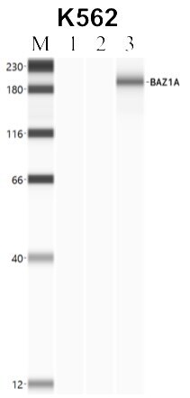
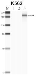
- Immunoprecipitation of BAZ1A was performed in K562 cells. Antigen-antibody complexes were formed by incubating approximately 500 µg whole cell lysate with 5 to 10 µL of polyclonal BAZ1A antibody (Product # PA5-51736) rotating 60 min at RT. The immune complexes were captured on 625 µg of anti-rabbit coated Dynabeads (Product # 11204D) and washed extensively. They were then eluted and analyzed using the Simple Western system using the same antibody as used in immunoprecipitation at a dilution of 1:25, followed by a 1:100 dilution of secondary antibody. Lane 1 is the input, lane 2 no antibody IP and lane 3 is the target specific IP. Data courtesy of the Yeo lab as part of the ENCODE project.

Supportive validation
- Submitted by
- Invitrogen Antibodies (provider)
- Main image
- Experimental details
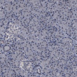
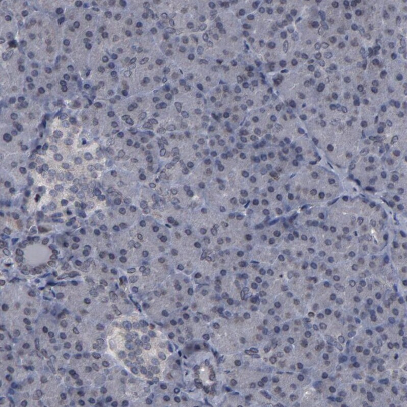
- Immunohistochemical analysis of BAZ1A in human pancreas using BAZ1A Polyclonal Antibody (Product # PA5-51736) shows no positivity in exocrine glandular cells as expected.
- Submitted by
- Invitrogen Antibodies (provider)
- Main image
- Experimental details
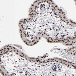
- Immunohistochemical analysis of BAZ1A in human placenta using BAZ1A Polyclonal Antibody (Product # PA5-51736) shows strong nuclear positivity in syncytiotrophoblast.

- Submitted by
- Invitrogen Antibodies (provider)
- Main image
- Experimental details
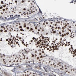
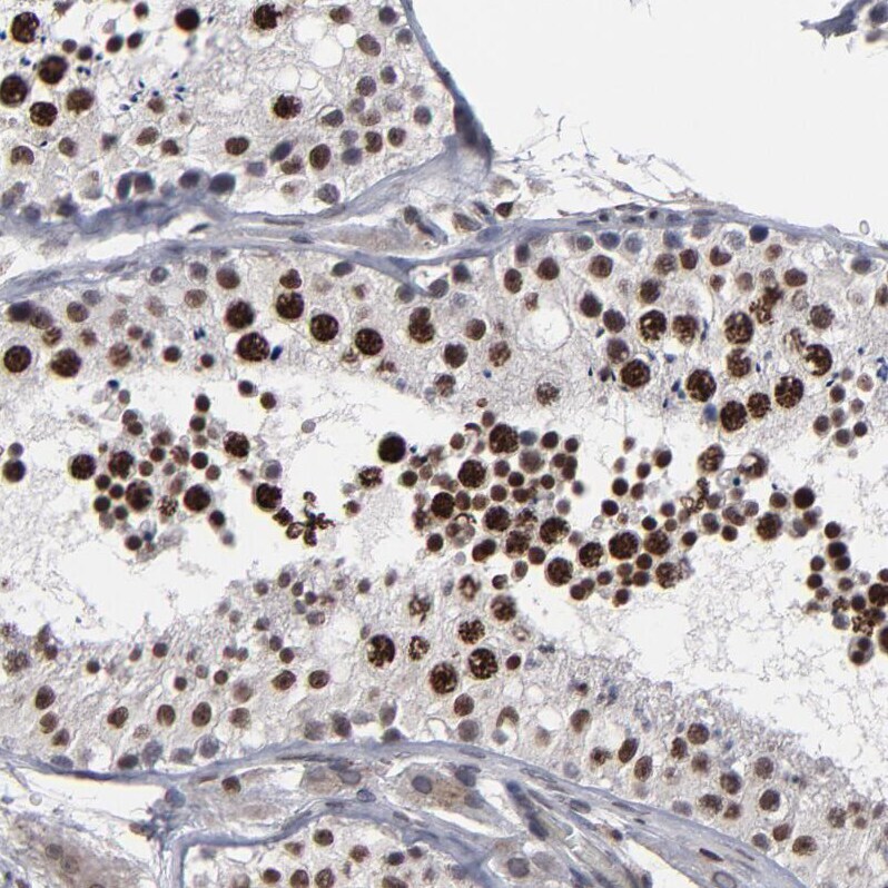
- Immunohistochemical analysis of BAZ1A in human testis using BAZ1A Polyclonal Antibody (Product # PA5-51736) shows strong nuclear positivity in cells in seminiferous ducts.
- Submitted by
- Invitrogen Antibodies (provider)
- Main image
- Experimental details
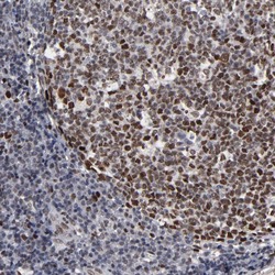
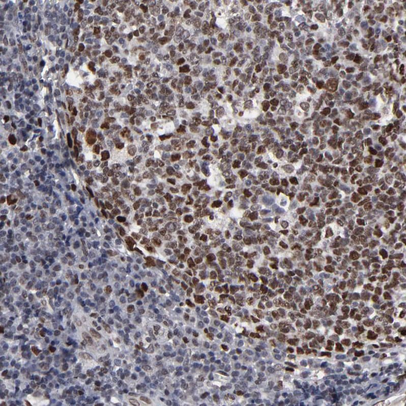
- Immunohistochemical analysis of BAZ1A in human tonsil using BAZ1A Polyclonal Antibody (Product # PA5-51736) shows strong nuclear positivity in germinal center cells.
Supportive validation
- Submitted by
- Invitrogen Antibodies (provider)
- Main image
- Experimental details
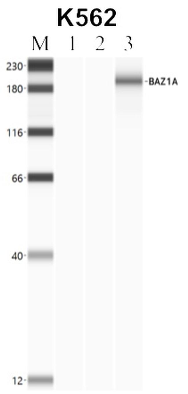
- Immunoprecipitation of BAZ1A was performed in K562 cells. Antigen-antibody complexes were formed by incubating approximately 500 µg whole cell lysate with 5 to 10 µL of polyclonal BAZ1A antibody (Product # PA5-51736) rotating 60 min at RT. The immune complexes were captured on 625 µg of anti-rabbit coated Dynabeads (Product # 11204D) and washed extensively. They were then eluted and analyzed using the Simple Western system using the same antibody as used in immunoprecipitation at a dilution of 1:25, followed by a 1:100 dilution of secondary antibody. Lane 1 is the input, lane 2 no antibody IP and lane 3 is the target specific IP. Data courtesy of the Yeo lab as part of the ENCODE project.
- Submitted by
- Invitrogen Antibodies (provider)
- Main image

- Experimental details
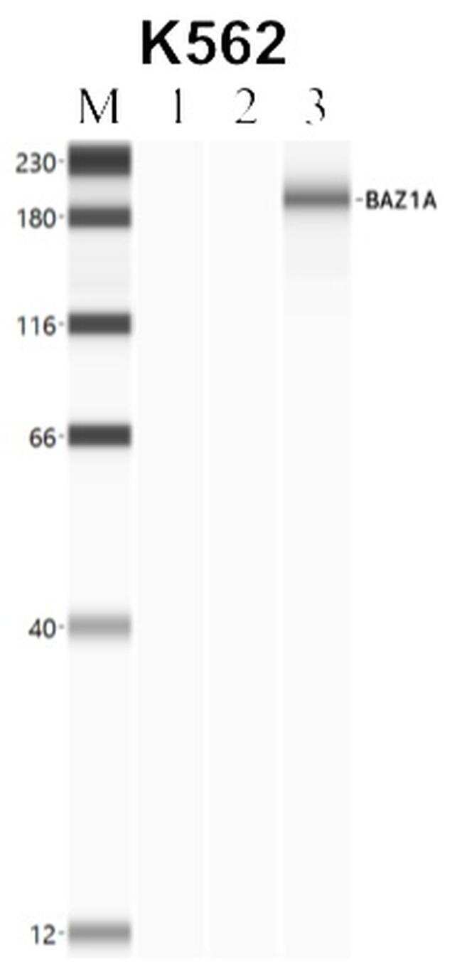
- Immunoprecipitation of BAZ1A was performed in K562 cells. Antigen-antibody complexes were formed by incubating approximately 500 µg whole cell lysate with 5 to 10 µL of polyclonal BAZ1A antibody (Product # PA5-51736) rotating 60 min at RT. The immune complexes were captured on 625 µg of anti-rabbit coated Dynabeads (Product # 11204D) and washed extensively. They were then eluted and analyzed using the Simple Western system using the same antibody as used in immunoprecipitation at a dilution of 1:25, followed by a 1:100 dilution of secondary antibody. Lane 1 is the input, lane 2 no antibody IP and lane 3 is the target specific IP. Data courtesy of the Yeo lab as part of the ENCODE project.
- Submitted by
- Invitrogen Antibodies (provider)
- Main image

- Experimental details
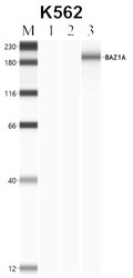
- RNA immunoprecipitation (RIP) western of BAZ1A was performed in K562 cells. Antigen-antibody complexes were formed by incubating approximately 500 µg whole cell lysate with 5 to 10 µL of polyclonal BAZ1A antibody (Product # PA5-51736) rotating 60 min at RT. The immune complexes were captured on 625 µg of anti-rabbit coated Dynabeads (Product # 11204D) and washed extensively. They were then eluted and analyzed using the Simple Western system using the same antibody as used in immunoprecipitation at a dilution of 1:25, followed by a 1:100 dilution of secondary antibody. Lane 1 is the input, lane 2 no antibody IP and lane 3 is the target specific IP. Data courtesy of the Yeo lab as part of the ENCODE project.

- Submitted by
- Invitrogen Antibodies (provider)
- Main image
- Experimental details
- RNA immunoprecipitation (RIP) western of BAZ1A was performed in K562 cells. Antigen-antibody complexes were formed by incubating approximately 500 µg whole cell lysate with 5 to 10 µL of polyclonal BAZ1A antibody (Product # PA5-51736) rotating 60 min at RT. The immune complexes were captured on 625 µg of anti-rabbit coated Dynabeads (Product # 11204D) and washed extensively. They were then eluted and analyzed using the Simple Western system using the same antibody as used in immunoprecipitation at a dilution of 1:25, followed by a 1:100 dilution of secondary antibody. Lane 1 is the input, lane 2 no antibody IP and lane 3 is the target specific IP. Data courtesy of the Yeo lab as part of the ENCODE project.