 Explore
 Validate
 Learn
Learn Western blot
Antibody data
- Antibody Data
- Antigen structure
- References [0]
- Comments [0]
- Validations
- Western blot [5]
- Immunohistochemistry [1]
Submit
Validation data
Reference
Comment
Report error
- Product number
- PA5-29256 - Provider product page

- Provider
- Invitrogen Antibodies
- Product name
- PAK1 Polyclonal Antibody
- Antibody type
- Polyclonal
- Antigen
- Recombinant full-length protein
- Description
- Recommended positive controls: A431, Raw264.7, PC-12. Predicted reactivity: Mouse (100%), Rat (100%), Zebrafish (98%), Xenopus laevis (95%), Chicken (99%), Bovine (100%). Store product as a concentrated solution. Centrifuge briefly prior to opening the vial.
- Reactivity
- Human, Mouse, Rat
- Host
- Rabbit
- Isotype
- IgG
- Vial size
- 100 μL
- Concentration
- 0.9 mg/mL
- Storage
- Store at 4°C short term. For long term storage, store at -20°C, avoiding freeze/thaw cycles.
No comments: Submit comment
Supportive validation
- Submitted by
- Invitrogen Antibodies (provider)
- Main image
- Experimental details
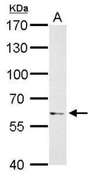
- PAK1 Polyclonal Antibody detects PAK1 protein by Western blot analysis. A. 30 µg PC-12 whole cell lysate/extract.7.5 % SDS-PAGE. PAK1 Polyclonal Antibody (Product # PA5-29256) dilution: 1:1,000.

- Submitted by
- Invitrogen Antibodies (provider)
- Main image

- Experimental details
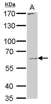
- PAK1 Polyclonal Antibody detects PAK1 protein by Western blot analysis. A. 30 µg Raw264.7 whole cell lysate/extract.7.5 % SDS-PAGE. PAK1 Polyclonal Antibody (Product # PA5-29256) dilution: 1:1,000.
- Submitted by
- Invitrogen Antibodies (provider)
- Main image
- Experimental details
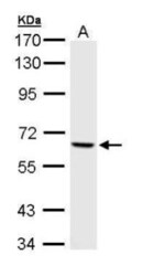
- Western Blot using PAK1 Polyclonal Antibody (Product # PA5-29256). Sample (30 µg of whole cell lysate). Lane A: A431 . 7.5% SDS PAGE. PAK1 Polyclonal Antibody (Product # PA5-29256) diluted at 1:1,000.

- Submitted by
- Invitrogen Antibodies (provider)
- Main image
- Experimental details
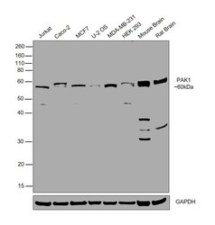
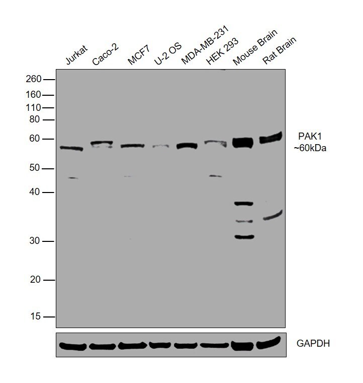
- Western blot was performed using Anti-PAK1 Polyclonal Antibody (Product # PA5-29256) and a 60kDa band corresponding to PAK1 was observed across all the tested cell lines and tissues. Whole cell extracts (30 µg lysate) of Jurkat (Lane 1), Caco-2 (Lane 2), MCF7 (Lane 3), U-2 OS (Lane 4), MDA-MB-231 (Lane 5), HEK-293 (Lane 6), Mouse Brain (Lane 7), Rat Brain (Lane 8) were electrophoresed using NuPAGE™ 10% Bis-Tris Protein Gel (Product # NP0302BOX). Resolved proteins were then transferred onto a Nitrocellulose membrane (Product # IB23001) by iBlot® 2 Dry Blotting System (Product # IB21001). The blot was probed with the primary antibody (1:1000 dilution) and detected by chemiluminescence with Goat anti-Rabbit IgG (Heavy Chain) Superclonal™ Recombinant Secondary Antibody, HRP (Product # A27036,1:4000) using the iBright FL 1000 (Product # A32752). Chemiluminescent detection was performed using SuperSignal™ West Dura Extended Duration Substrate (Product # 34076).
- Submitted by
- Invitrogen Antibodies (provider)
- Main image
- Experimental details
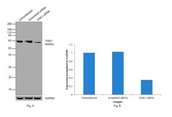
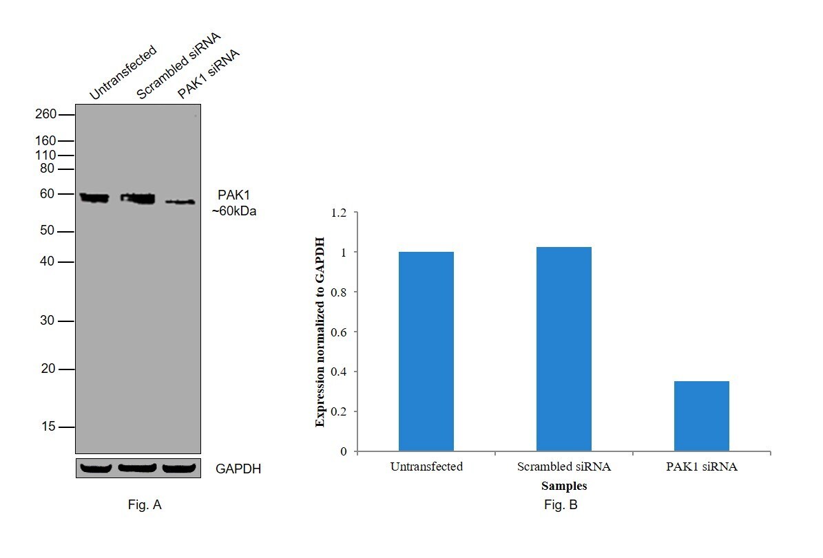
- Knockdown of PAK1 was achieved by transfecting Caco-2 with PAK1 specific siRNAs (Silencer® select Product # S10020, S10021). Western blot analysis (Fig. A) was performed using Whole cell extracts from the PAK1 knockdown cells (lane 3), non-targeting scrambled siRNA transfected cells (lane 2) and untransfected cells (lane 1). The blot was probed with PAK1 Polyclonal Antibody (Product # PA5-29256, 1:1000 dilution) and Goat anti-Rabbit IgG (Heavy Chain) Superclonal™ Recombinant Secondary Antibody, HRP (Product # A27036, 1:4000). Densitometric analysis of this western blot is shown in histogram (Fig. B). Decrease in signal upon siRNA mediated knock down confirms that antibody is specific to PAK1.
Supportive validation
- Submitted by
- Invitrogen Antibodies (provider)
- Main image
- Experimental details
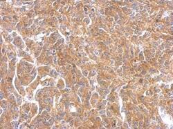
- PAK1 Polyclonal Antibody detects PAK1 protein at on Saos2 xenograft by immunohistochemical analysis. Sample: Paraffin-embedded Saos2 xenograft. PAK1 Polyclonal Antibody (Product # PA5-29256) dilution: 1:500. Antigen Retrieval: EDTA based buffer, pH 8.0, 15 min.
