Explore
 Validate
 Learn
Learn Western blot
 Immunocytochemistry
Antibody data
- Antibody Data
- Antigen structure
- References [2]
- Comments [0]
- Validations
- Immunocytochemistry [1]
- Immunohistochemistry [1]
Submit
Validation data
Reference
Comment
Report error
- Product number
- HPA003565 - Provider product page

- Provider
- Atlas Antibodies
- Proper citation
- Atlas Antibodies Cat#HPA003565, RRID:AB_1854935
- Product name
- Anti-PAK1
- Antibody type
- Polyclonal
- Description
- Polyclonal Antibody against Human PAK1, Gene description: p21 protein (Cdc42/Rac)-activated kinase 1, Validated applications: ICC, WB, IHC, Uniprot ID: Q13153, Storage: Store at +4°C for short term storage. Long time storage is recommended at -20°C.
- Reactivity
- Human, Mouse, Rat
- Host
- Rabbit
- Conjugate
- Unconjugated
- Isotype
- IgG
- Vial size
- 100 µl
- Concentration
- 0.2 mg/ml
- Storage
- Store at +4°C for short term storage. Long time storage is recommended at -20°C.
- Handling
- The antibody solution should be gently mixed before use.
Submitted references A requirement for PAK1 to support mitochondrial function and maintain cellular redox balance via electron transport chain proteins to prevent β-cell apoptosis
Initial Quantitative Proteomic Map of 28 Mouse Tissues Using the SILAC Mouse
Ahn M, Oh E, McCown E, Wang X, Veluthakal R, Thurmond D
Metabolism 2021;115
Metabolism 2021;115
Initial Quantitative Proteomic Map of 28 Mouse Tissues Using the SILAC Mouse
Geiger T, Velic A, Macek B, Lundberg E, Kampf C, Nagaraj N, Uhlen M, Cox J, Mann M
Molecular & Cellular Proteomics 2013;12(6):1709-1722
Molecular & Cellular Proteomics 2013;12(6):1709-1722
No comments: Submit comment
Supportive validation
- Submitted by
- Atlas Antibodies (provider)
- Main image

- Experimental details
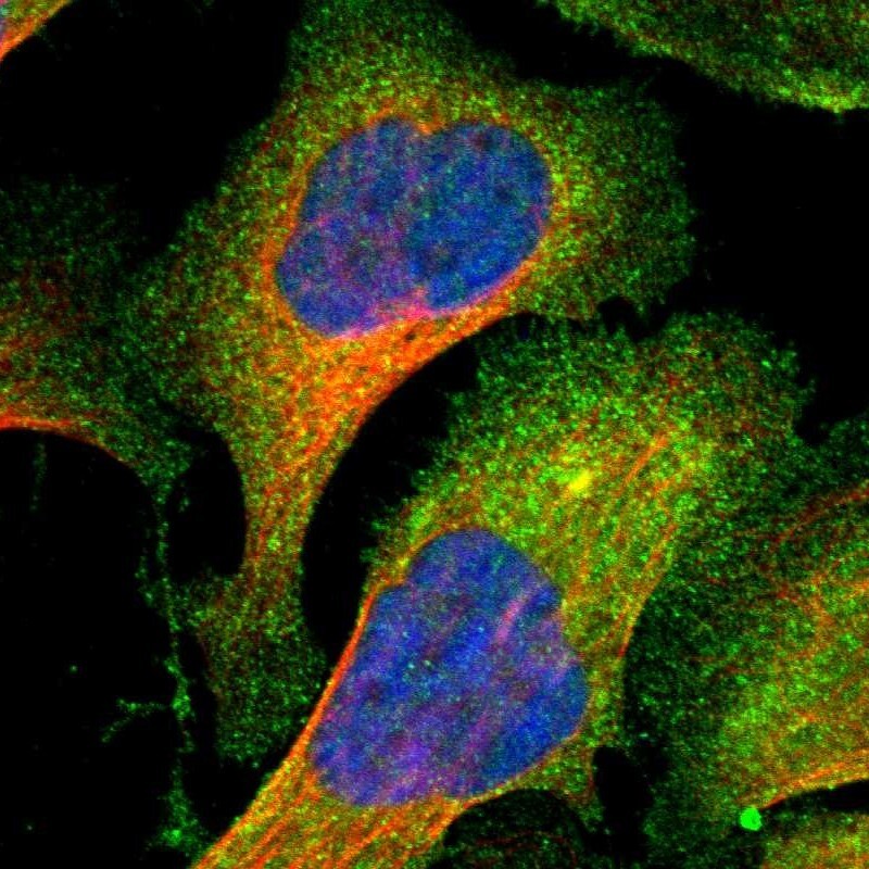
- Immunofluorescent staining of human cell line U-2 OS shows localization to plasma membrane & cytosol.
- Sample type
- Human
Supportive validation
- Submitted by
- Atlas Antibodies (provider)
- Enhanced method
- Orthogonal validation
- Main image
- Experimental details
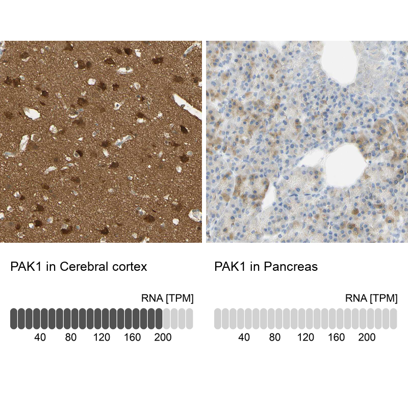
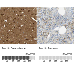
- Immunohistochemistry analysis in human cerebral cortex and pancreas tissues using HPA003565 antibody. Corresponding PAK1 RNA-seq data are presented for the same tissues.
- Sample type
- Human
- Protocol
- Protocol