Explore
 Validate
 Learn
Learn Western blot
 Immunocytochemistry
Antibody data
- Antibody Data
- Antigen structure
- References [4]
- Comments [0]
- Validations
- Immunocytochemistry [4]
Submit
Validation data
Reference
Comment
Report error
- Product number
- PA1-978 - Provider product page

- Provider
- Invitrogen Antibodies
- Product name
- PSMB6 Polyclonal Antibody
- Antibody type
- Polyclonal
- Antigen
- Synthetic peptide
- Description
- PA1-978 detects proteasome 20S Y from purified bovine and human 26S proteasome samples. PA1-978 has been successfully used in Western blot and ICC/IF procedures. By Western blot, this antibody detects an ~25 kDa protein representing proteasome 20S Y from purified human 26S proteasome. The PA1-978 immunizing peptides correspond to amino acid residues 41-58 from human proteasome 20S Y and amino acid residues 74-91 from frog. These sequences are completely conserved in mouse, rat, and zebrafish. PA1-978 immunizing peptide (Cat. # PEP-173) is available for use in neutralization and control procedures.
- Reactivity
- Human, Mouse, Rat, Bovine
- Host
- Rabbit
- Isotype
- IgG
- Vial size
- 100 µg
- Concentration
- 1 mg/mL
- Storage
- -20° C, Avoid Freeze/Thaw Cycles
Submitted references Evaluation of Immunoproteasome-Specific Proteolytic Activity Using Fluorogenic Peptide Substrates.
The ubiquitin-like protein FAT10 mediates NF-kappaB activation.
Persistent hijacking of brain proteasomes in HIV-associated dementia.
Reduced ubiquitin C-terminal hydrolase-1 expression levels in dementia with Lewy bodies.
Kim S, Park SH, Choi WH, Lee MJ
Immune network 2022 Jun;22(3):e28
Immune network 2022 Jun;22(3):e28
The ubiquitin-like protein FAT10 mediates NF-kappaB activation.
Gong P, Canaan A, Wang B, Leventhal J, Snyder A, Nair V, Cohen CD, Kretzler M, D'Agati V, Weissman S, Ross MJ
Journal of the American Society of Nephrology : JASN 2010 Feb;21(2):316-26
Journal of the American Society of Nephrology : JASN 2010 Feb;21(2):316-26
Persistent hijacking of brain proteasomes in HIV-associated dementia.
Nguyen TP, Soukup VM, Gelman BB
The American journal of pathology 2010 Feb;176(2):893-902
The American journal of pathology 2010 Feb;176(2):893-902
Reduced ubiquitin C-terminal hydrolase-1 expression levels in dementia with Lewy bodies.
Barrachina M, Castaño E, Dalfó E, Maes T, Buesa C, Ferrer I
Neurobiology of disease 2006 May;22(2):265-73
Neurobiology of disease 2006 May;22(2):265-73
No comments: Submit comment
Supportive validation
- Submitted by
- Invitrogen Antibodies (provider)
- Main image
- Experimental details
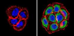
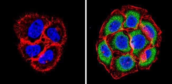
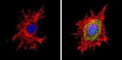
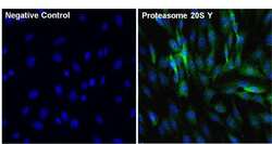
- Immunofluorescent analysis of Proteasome 20S Y (green) showing staining in the cytoplasm and nucleus of A431 cells (right) compared to a negative control without primary antibody (left). Formalin-fixed cells were permeabilized with 0.1% Triton X-100 in TBS for 5-10 minutes and blocked with 3% BSA-PBS for 30 minutes at room temperature. Cells were probed with a Proteasome 20S Y polyclonal antibody (Product # PA1-978) in 3% BSA-PBS at a dilution of 1:20 and incubated overnight at 4 ºC in a humidified chamber. Cells were washed with PBST and incubated with a DyLight-conjugated secondary antibody in PBS at room temperature in the dark. F-actin (red) was stained with a fluorescent red phalloidin and nuclei (blue) were stained with Hoechst or DAPI. Images were taken at a magnification of 60x.
- Submitted by
- Invitrogen Antibodies (provider)
- Main image
- Experimental details
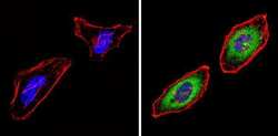
- Immunofluorescent analysis of Proteasome 20S Y (green) showing staining in the cytoplasm and nucleus of BAEC cells (right) compared to a negative control without primary antibody (left). Formalin-fixed cells were permeabilized with 0.1% Triton X-100 in TBS for 5-10 minutes and blocked with 3% BSA-PBS for 30 minutes at room temperature. Cells were probed with a Proteasome 20S Y polyclonal antibody (Product # PA1-978) in 3% BSA-PBS at a dilution of 1:20 and incubated overnight at 4 ºC in a humidified chamber. Cells were washed with PBST and incubated with a DyLight-conjugated secondary antibody in PBS at room temperature in the dark. F-actin (red) was stained with a fluorescent red phalloidin and nuclei (blue) were stained with Hoechst or DAPI. Images were taken at a magnification of 60x.
- Submitted by
- Invitrogen Antibodies (provider)
- Main image
- Experimental details
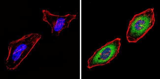
- Immunofluorescent analysis of Proteasome 20S Y (green) showing staining in the cytoplasm and nucleus of Hela cells (right) compared to a negative control without primary antibody (left). Formalin-fixed cells were permeabilized with 0.1% Triton X-100 in TBS for 5-10 minutes and blocked with 3% BSA-PBS for 30 minutes at room temperature. Cells were probed with a Proteasome 20S Y polyclonal antibody (Product # PA1-978) in 3% BSA-PBS at a dilution of 1:20 and incubated overnight at 4 ºC in a humidified chamber. Cells were washed with PBST and incubated with a DyLight-conjugated secondary antibody in PBS at room temperature in the dark. F-actin (red) was stained with a fluorescent red phalloidin and nuclei (blue) were stained with Hoechst or DAPI. Images were taken at a magnification of 60x.
- Submitted by
- Invitrogen Antibodies (provider)
- Main image
- Experimental details
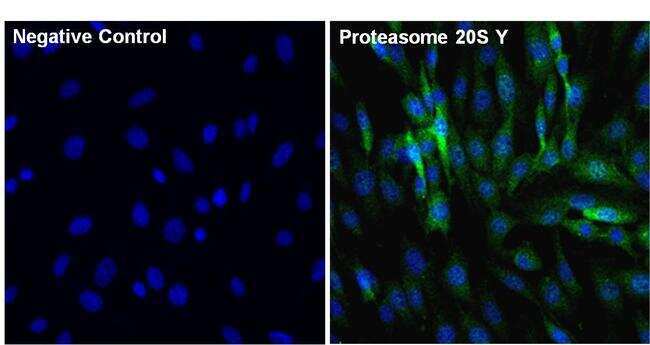
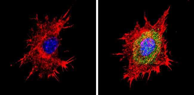
- Immunofluorescent analysis of Proteasome 20S Y (green) in 3T3 cells. The cells were fixed with 4% paraformaldehyde for 15 minutes, permeabilized with 0.1% Triton X-100 in PBS for 15 minutes and blocked with 3% Blocker BSA (Product # 37525) in PBS for 30 minutes at room temperature. Cells were stained with or without Proteasome 20S Y rabbit polyclonal antibody (Product # PA1-978), at a concentration of 5 µg/mL for 1 hour at room temperature, and then incubated with a Goat anti-Rabbit (H+L) Superclonal Secondary Antibody, Alexa Fluor® 488 conjugate (Product # A27034) at a dilution of 1:1000 for 1 hour at room temperature (both panels, green). Nuclei (both panels, blue) were stained with Hoechst 33342 dye (Product # 62249). Images were taken on a Thermo Scientific ToxInsight at 20X magnification.