Explore
 Validate
 Learn
Learn Western blot
 Immunoprecipitation
Antibody data
- Antibody Data
- Antigen structure
- References [10]
- Comments [0]
- Validations
- Western blot [3]
- Other assay [3]
Submit
Validation data
Reference
Comment
Report error
- Product number
- 44-686G - Provider product page

- Provider
- Invitrogen Antibodies
- Product name
- Phospho-CDK1 (Thr14, Tyr15) Polyclonal Antibody
- Antibody type
- Polyclonal
- Antigen
- Synthetic peptide
- Reactivity
- Human, Mouse, Rat
- Host
- Rabbit
- Isotype
- IgG
- Vial size
- 100 µL
- Storage
- -20°C
Submitted references Mre11 exonuclease activity promotes irreversible mitotic progression under replication stress.
Ongoing replication forks delay the nuclear envelope breakdown upon mitotic entry.
Metabolite and thymocyte development defects in ADA-SCID mice receiving enzyme replacement therapy.
Effects of Oncogenic Gα(q) and Gα(11) Inhibition by FR900359 in Uveal Melanoma.
4-Hydroxybenzoic acid derivatives as HDAC6-specific inhibitors modulating microtubular structure and HSP90α chaperone activity against prostate cancer.
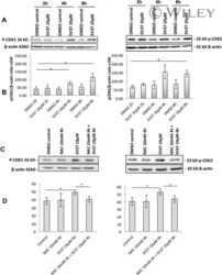
A novel coumarin-quinone derivative SV37 inhibits CDC25 phosphatases, impairs proliferation, and induces cell death.
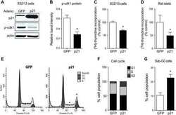
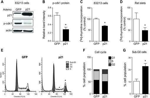
Upregulation of p21 activates the intrinsic apoptotic pathway in β-cells.
Multisite phosphoregulation of Cdc25 activity refines the mitotic entrance and exit switches.
The extracellular signal-regulated kinase-mitogen-activated protein kinase pathway phosphorylates and targets Cdc25A for SCF beta-TrCP-dependent degradation for cell cycle arrest.
Dynamic expression of the RNA-binding protein Sam68 during mouse pre-implantation development.
Hashimoto Y, Tanaka H
Life science alliance 2022 Jun;5(6)
Life science alliance 2022 Jun;5(6)
Ongoing replication forks delay the nuclear envelope breakdown upon mitotic entry.
Hashimoto Y, Tanaka H
The Journal of biological chemistry 2021 Jan-Jun;296:100033
The Journal of biological chemistry 2021 Jan-Jun;296:100033
Metabolite and thymocyte development defects in ADA-SCID mice receiving enzyme replacement therapy.
Moretti FA, Giardino G, Attenborough TCH, Gkazi AS, Margetts BK, la Marca G, Fairbanks L, Crompton T, Gaspar HB
Scientific reports 2021 Dec 1;11(1):23221
Scientific reports 2021 Dec 1;11(1):23221
Effects of Oncogenic Gα(q) and Gα(11) Inhibition by FR900359 in Uveal Melanoma.
Lapadula D, Farias E, Randolph CE, Purwin TJ, McGrath D, Charpentier TH, Zhang L, Wu S, Terai M, Sato T, Tall GG, Zhou N, Wedegaertner PB, Aplin AE, Aguirre-Ghiso J, Benovic JL
Molecular cancer research : MCR 2019 Apr;17(4):963-973
Molecular cancer research : MCR 2019 Apr;17(4):963-973
4-Hydroxybenzoic acid derivatives as HDAC6-specific inhibitors modulating microtubular structure and HSP90α chaperone activity against prostate cancer.
Seidel C, Schnekenburger M, Mazumder A, Teiten MH, Kirsch G, Dicato M, Diederich M
Biochemical pharmacology 2016 Jan 1;99:31-52
Biochemical pharmacology 2016 Jan 1;99:31-52
A novel coumarin-quinone derivative SV37 inhibits CDC25 phosphatases, impairs proliferation, and induces cell death.
Bana E, Sibille E, Valente S, Cerella C, Chaimbault P, Kirsch G, Dicato M, Diederich M, Bagrel D
Molecular carcinogenesis 2015 Mar;54(3):229-41
Molecular carcinogenesis 2015 Mar;54(3):229-41
Upregulation of p21 activates the intrinsic apoptotic pathway in β-cells.
Hernandez AM, Colvin ES, Chen YC, Geiss SL, Eller LE, Fueger PT
American journal of physiology. Endocrinology and metabolism 2013 Jun 15;304(12):E1281-90
American journal of physiology. Endocrinology and metabolism 2013 Jun 15;304(12):E1281-90
Multisite phosphoregulation of Cdc25 activity refines the mitotic entrance and exit switches.
Lu LX, Domingo-Sananes MR, Huzarska M, Novak B, Gould KL
Proceedings of the National Academy of Sciences of the United States of America 2012 Jun 19;109(25):9899-904
Proceedings of the National Academy of Sciences of the United States of America 2012 Jun 19;109(25):9899-904
The extracellular signal-regulated kinase-mitogen-activated protein kinase pathway phosphorylates and targets Cdc25A for SCF beta-TrCP-dependent degradation for cell cycle arrest.
Isoda M, Kanemori Y, Nakajo N, Uchida S, Yamashita K, Ueno H, Sagata N
Molecular biology of the cell 2009 Apr;20(8):2186-95
Molecular biology of the cell 2009 Apr;20(8):2186-95
Dynamic expression of the RNA-binding protein Sam68 during mouse pre-implantation development.
Paronetto MP, Bianchi E, Geremia R, Sette C
Gene expression patterns : GEP 2008 May;8(5):311-22
Gene expression patterns : GEP 2008 May;8(5):311-22
No comments: Submit comment
Supportive validation
- Submitted by
- Invitrogen Antibodies (provider)
- Main image

- Experimental details
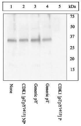
- Peptide Competition: Extracts prepared from HeLa cells stimulated with nocozadole were resolved by SDS-PAGE on a 10% polyacrylamide gel and transferred to PVDF. Membranes were blocked with a 5% BSA-TBST buffer for two hours at room temperature, then were
- Submitted by
- Invitrogen Antibodies (provider)
- Main image
- Experimental details
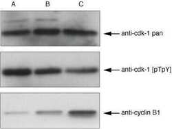
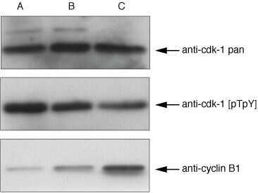
- cdk1 antibodyy (Product # AHZ0112) and cdk1 (pTpY14⁄15) phosphospecific antibody. Western blots were incubated with cdk1, cdk1 (pTpY), and cyclin B1 (Product # AHF0062) antibodies. Jurkat cells were a) serum starved, b) proliferating, and c) treated with nocozadole, each for 18 hours.
- Submitted by
- Invitrogen Antibodies (provider)
- Main image

- Experimental details
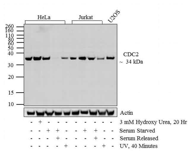
- Western blot analysis was performed on whole cell extracts (30 µg lysate) of HeLa (1), HeLa treated for 20 hr with 3mM of Hydroxy Urea (2), HeLa Serum Starved (3), HeLa Serum Starved overnight following by serum release (4), HeLa treated with UV for 40 min (5), Jurkat (6), Jurkat Serum Starved (7), Jurkat Serum Starved overnight following by serum release (8), Jurkat treated with UV for 40 min (9) and U2OS (10). The blots were probed with Anti-CDC2 (pT14/pY15) Rabbit Polyclonal Antibody (Product # 44-686G, 1:500-1:2000 µg/mL) and detected by chemiluminescence using Goat anti-Rabbit IgG (H+L) Secondary Antibody, HRP conjugate (Product # G-21234, 1:5000 dilution). A 34 kDa band corresponding to CDC2 (pT14/pY15) was observed across cell lines tested. Upon UV treatment and Serum starvation overnight followed by serum release, the expression of CDC2 (pT14/pY15) was decreased. Known quantity of protein samples were electrophoresed using Novex® NuPAGE® 12% Bis-Tris gel (Product # NP0342BOX), XCell SureLock™ Electrophoresis System (Product # EI0002) and Novex® Sharp Pre-Stained Protein Standard (Product # LC5800). Resolved proteins were then transferred onto a transferred onto a nitrocellulose membrane with iBlot® 2 Dry Blotting System (Product # IB21001). The membrane was probed with the relevant primary and secondary Antibody following blocking with 5 % skimmed milk. Chemiluminescent detection was performed using Novex® ECL Chemiluminescent Substrate Reage
Supportive validation
- Submitted by
- Invitrogen Antibodies (provider)
- Main image
- Experimental details
- NULL
- Submitted by
- Invitrogen Antibodies (provider)
- Main image
- Experimental details
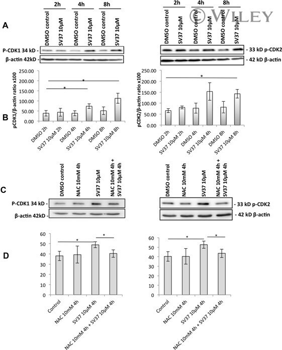
- (A) Analysis of pCDK1 and pCDK2 levels in MDA-MB-231 cells treated with SV37 at 10 uM or untreated, for different treatment times, with (B) corresponding ratios of pCDK1/beta-actin and pCDK2/beta-actin. Significance was calculated for each assay versus corresponding DMSO control (values were calculated from quantification of Western blot signal). Results show an increase of CDK1 and CDK2 phosphorylation after 4 h of treatment with SV37 10 uM. (C) Analysis of pCDK1 and pCDK2 levels in MDA-MB-231 cells pretreated with NAC at 10 mM and treated by SV37 at 10 uM or untreated, for different treatment times with (D) corresponding ratios of pCDK1/beta-actin and pCDK2/beta-actin.
- Submitted by
- Invitrogen Antibodies (provider)
- Main image

- Experimental details
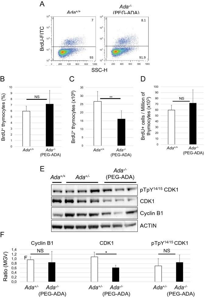
- Figure 7 Ado accumulation does not interfere with thymocytes proliferation. ( A ) FACS plots of proliferating thymocytes (BrdU + ) from control ( Ada + / + ) and 3-week-PEG-ADA-treated Ada -/- mice. Bar graphs representing percentages ( B ), absolute ( C ) and relative ( D ) numbers of BrdU + thymocytes shown in (A) (n = 6, 6). ( E ) Western blot analysis of protein lysates of thymocytes shown in (A). Anti-ACTIN stain was used as protein loading control. Full-length blots are presented in Supplementary Fig. 12 . ( F ) ImageJ quantification of protein bands from Western blot film shown in (E). MGV = mean gray value. * P < 0.05, ** P < 0.01 and *** P < 0.001. NS = statistically not significant.