Explore
 Validate
 Learn
Learn Western blot
Antibody data
- Antibody Data
- Antigen structure
- References [1]
- Comments [0]
- Validations
- Western blot [1]
- Immunocytochemistry [1]
- Flow cytometry [1]
- Other assay [1]
Submit
Validation data
Reference
Comment
Report error
- Product number
- 701808 - Provider product page

- Provider
- Invitrogen Antibodies
- Product name
- Phospho-CDK1 (Thr14, Tyr15) Recombinant Rabbit Monoclonal Antibody (17H29L7)
- Antibody type
- Monoclonal
- Antigen
- Synthetic peptide
- Reactivity
- Human
- Host
- Rabbit
- Isotype
- IgG
- Antibody clone number
- 17H29L7
- Vial size
- 100 µg
- Concentration
- 0.5 mg/mL
- Storage
- Store at 4°C short term. For long term storage, store at -20°C, avoiding freeze/thaw cycles.
Submitted references EXOSC10 sculpts the transcriptome during the growth-to-maturation transition in mouse oocytes.
Wu D, Dean J
Nucleic acids research 2020 Jun 4;48(10):5349-5365
Nucleic acids research 2020 Jun 4;48(10):5349-5365
No comments: Submit comment
Supportive validation
- Submitted by
- Invitrogen Antibodies (provider)
- Main image

- Experimental details
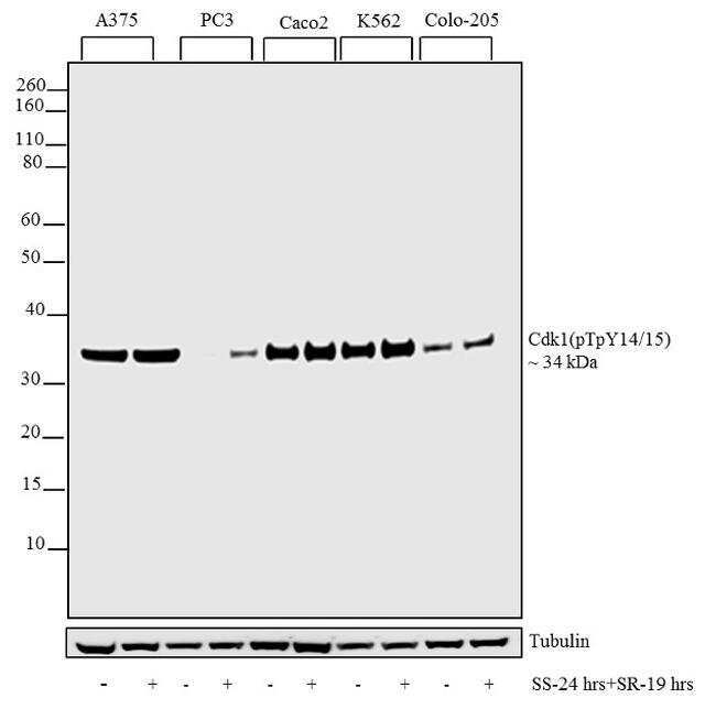
- Western blot analysis was performed on whole cell extracts (30 µg lysate) of A375 (1), A375 (Serum starved24 hours and released for 19 hours) (2), PC3 (3), PC3 (Serum starved24 hours and released for 19 hours) (4), Caco2 (5), Caco2 (Serum starved24 hours and released for 19 hours) (6), K562 (7), K562 (Serum starved24 hours and released for 19 hours) (8), Colo-205 (9) and Colo-205 (Serum starved24 hours and released for 19 hours) (10). The blots were probed with Anti-Cdk1 (pTpY14/15) Recombinant Rabbit Monoclonal Antibody (Product # 701808, 1-2 µg/mL) and detected by chemiluminescence using Goat anti-Rabbit IgG (H+L) Superclonal™ Secondary Antibody, HRP conjugate (Product # A27036, 0.4 µg/mL, 1:2500 dilution). A 34 kDa band corresponding to Cdk1 (pTpY14/15) was observed across cell lines tested. Treatment response is more evident in cell lines where basal expression is less. Known quantity of protein samples were electrophoresed using Novex® NuPAGE® 4-12% Bis-Tris gel (Product # NP0321BOX), XCell SureLock™ Electrophoresis System (Product # EI0002), and Novex® Sharp Pre-Stained Protein Standard (Product # LC5800). Resolved proteins were then transferred onto a nitrocellulose membrane with iBlot® Dry Blotting System (Product # IB21001). The membrane was probed with the relevant primary and secondary Antibody following blocking with 5% skimmed milk. Chemiluminescent detection was performed using Novex® ECL Chemiluminescent Substrate
Supportive validation
- Submitted by
- Invitrogen Antibodies (provider)
- Main image
- Experimental details
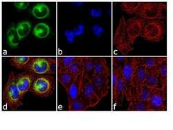
- Immunofluorescence was performed on fixed and permeabilized A375 cells which were serum starved (19 hours) and serum released (24 hours), for detection of Cdk1 (pTpY14/15) using Anti- Cdk1 (pTpY14/15) Recombinant Rabbit Monoclonal Antibody (Product # 701808, 1-2 µg/mL) and labeled with Goat anti-Rabbit IgG (H+L) Superclonal™ Secondary Antibody, Alexa Fluor® 488 conjugate (Product # A27034, 1:2000). Panel a) shows representative cells that were stained for detection and localization of Cdk1 (pTpY14/15) protein (green), Panel b) is stained for nuclei (blue) using SlowFade® Gold Antifade Mountant with DAPI (Product # S36938). Panel c) represents cytoskeletal F-actin staining using Alexa Fluor® 555 Rhodamine Phalloidin (Product # R415, 1:300). Panel d) is a composite image of Panels a, b and c clearly demonstrating cytoplasmic localization of Cdk1 (pTpY14/15) Panel e) shows no signal which demonstrates antibody specificity against Cdk1 (pTpY14/15) phosphorylated peptide (Antibody was incubated with phosphorylated peptide for 1 hour/37C). Panel f) represents control cells with no primary Antibody to assess background.
Supportive validation
- Submitted by
- Invitrogen Antibodies (provider)
- Main image

- Experimental details
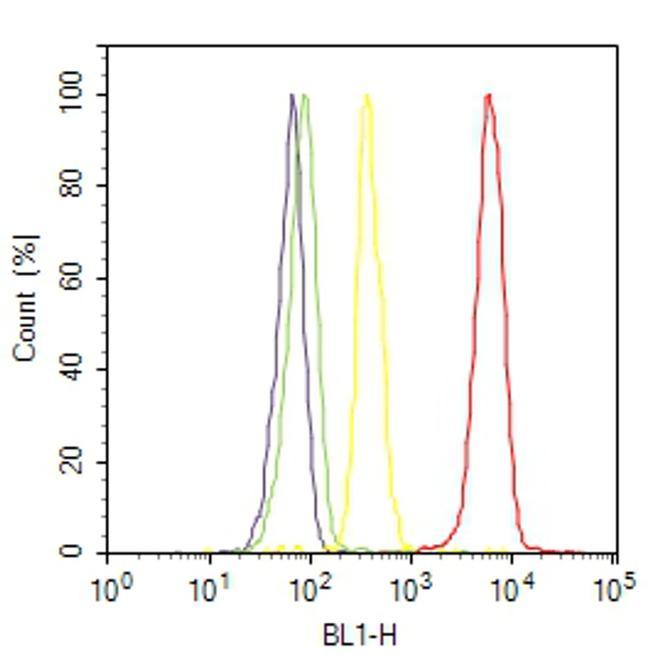
- Flow Cytometry analysis of Cdk1 [pT14/pY15] was performed on K-562 cells (serum starved overnight followed by serum release for 4 hours) labeled with ABfinity™ Anti-Cdk1 [pT14/pY15] Recombinant Rabbit Monoclonal Antibody (Product# 701808, 2-4 ug/ 1M cells) or with Rabbit isotype control and detected with Goat anti-Rabbit IgG (H+L) Superclonal™ Secondary Antibody, (Alexa Fluor® 488 conjugate, Product # A27034, 0.4 ug/ml, 1:2500) as represented by the red and yellow histograms respectively. The purple histogram represents unstained control cells and the green histogram represents no-primary-Antibody control. A representative of 10,000 cells were acquired and analyzed for each sample using an Attune® Acoustic Focusing Cytometer (4468770).
Supportive validation
- Submitted by
- Invitrogen Antibodies (provider)
- Main image

- Experimental details
- NULL