Explore
 Validate
 Learn
Learn Western blot
Antibody data
- Antibody Data
- Antigen structure
- References [2]
- Comments [0]
- Validations
- Western blot [1]
- Immunocytochemistry [1]
- Other assay [1]
Submit
Validation data
Reference
Comment
Report error
- Product number
- 710840 - Provider product page

- Provider
- Invitrogen Antibodies
- Product name
- Phospho-CDK1 (Thr14, Tyr15) Recombinant Polyclonal Antibody (17 HCLC)
- Antibody type
- Polyclonal
- Antigen
- Synthetic peptide
- Description
- This antibody is predicted to react with Monkey, Rat, Mouse, Rabbit, Bovine, Pig and Feline.
- Antibody clone number
- 17 HCLC
- Concentration
- 0.5 mg/mL
Submitted references In Silico Identification of Small Molecules as New Cdc25 Inhibitors through the Correlation between Chemosensitivity and Protein Expression Pattern.
The Tumor Suppressor MIG6 Controls Mitotic Progression and the G2/M DNA Damage Checkpoint by Stabilizing the WEE1 Kinase.
Lauria A, Martorana A, La Monica G, Mannino S, Mannino G, Peri D, Gentile C
International journal of molecular sciences 2021 Apr 2;22(7)
International journal of molecular sciences 2021 Apr 2;22(7)
The Tumor Suppressor MIG6 Controls Mitotic Progression and the G2/M DNA Damage Checkpoint by Stabilizing the WEE1 Kinase.
Sasaki M, Terabayashi T, Weiss SM, Ferby I
Cell reports 2018 Jul 31;24(5):1278-1289
Cell reports 2018 Jul 31;24(5):1278-1289
No comments: Submit comment
Supportive validation
- Submitted by
- Invitrogen Antibodies (provider)
- Main image

- Experimental details
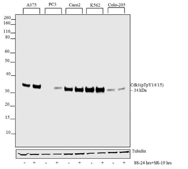
- Western blot analysis was performed on whole cell extracts (30 µg lysate) of A375 (1), A375 (Serum starved24 hours and released for 19 hours) (2), PC3 (3), PC3 (Serum starved24 hours and released for 19 hours) (4), Caco2 (5), Caco2 (Serum starved24 hours and released for 19 hours) (6), K562 (7), K562 (Serum starved24 hours and released for 19 hours) (8), Colo-205 (9) and Colo-205 (Serum starved24 hours and released for 19 hours) (10). The blots were probed with Anti-Cdk1 (pTpY14/15) Recombinant Rabbit Polyclonal Antibody (Product # 710840, 1-2 µg/mL) and detected by chemiluminescence using Goat anti-Rabbit IgG (H+L) Superclonal™ Secondary Antibody, HRP conjugate (Product # A27036, 0.4 µg/mL, 1:2500 dilution). A 34 kDa band corresponding to Cdk1 (pTpY14/15) was observed across cell lines tested. Treatment response is more evident in cell lines where basal expression is less. Known quantity of protein samples were electrophoresed using Novex® NuPAGE® 4-12% Bis-Tris gel (Product # NP0321BOX), XCell SureLock™ Electrophoresis System (Product # EI0002), and Novex® Sharp Pre-Stained Protein Standard (Product # LC5800). Resolved proteins were then transferred onto a nitrocellulose membrane with iBlot® Dry Blotting System (Product # IB21001). The membrane was probed with the relevant primary and secondary Antibody following blocking with 5% skimmed milk. Chemiluminescent detection was performed using Novex® ECL Chemiluminescent Substrat
Supportive validation
- Submitted by
- Invitrogen Antibodies (provider)
- Main image

- Experimental details
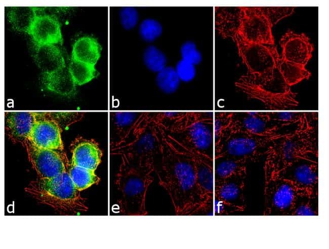
- Immunofluorescence was performed on fixed and permeabilized A375 cells which were serum starved (19 hours) and serum released (24 hours) for detection of Cdk1 (pTpY14/15) using Anti-Cdk1 (pTpY14/15) Recombinant Rabbit Polyclonal Antibody (Product # 710840, 1-2 µg/mL) and labeled with Goat anti-Rabbit IgG (H+L) Superclonal™ Secondary Antibody, Alexa Fluor® 488 conjugate (Product # A27034, 1:2000). Panel a) shows representative cells that were stained for detection and localization of Cdk1 (pTpY14/15) protein (green), Panel b) is stained for nuclei (blue) using SlowFade® Gold Antifade Mountant with DAPI (Product # S36938). Panel c) represents cytoskeletal F-actin staining using Alexa Fluor® 555 Rhodamine Phalloidin (Product # R415, 1:300). Panel d) is a composite image of Panels a, b and c clearly demonstrating cytoplasmic localization of Cdk1 (pTpY14/15) Panel e) shows no signal which demonstrates antibody specificity against Cdk1 (pTpY14/15) phosphorylated peptide (Antibody was incubated with phosphorylated peptide for 1 hour/37C). Panel f) represents control cells with no primary Antibody to assess background.
Supportive validation
- Submitted by
- Invitrogen Antibodies (provider)
- Main image
- Experimental details
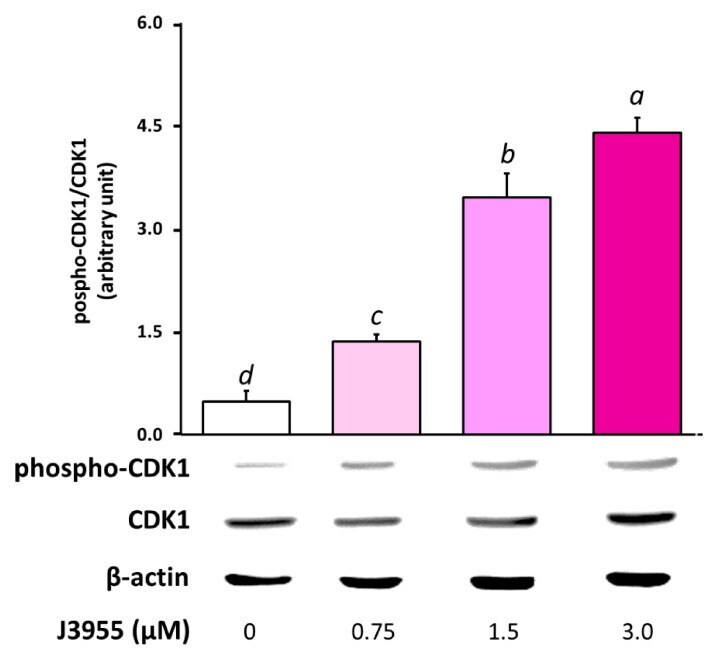
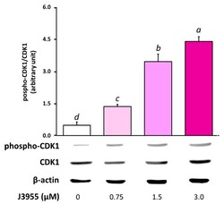
- Figure 10 Effects of a 6 h treatment with J3955 (0.75, 1.5, and 3 muM) on Cdk1 phosphorylation in HepG2 cells. After the treatment, cells were collected and the proteins were isolated for Western blot analysis as described in ''''Materials and methods''''. The panel shows a representative Western blot and densitometric analysis. The values represent the ratio between phospho-Cdk1 and total Cdk1, both previously normalized for the corresponding beta-actin. Values are expressed as the mean +- S.D. of three separate experiments with similar results. Different lowercase letters on the top of each histogram indicate statistical ( p < 0.05) differences among the tested samples, as measured by one-way ANOVA followed by the Tuckey test. The letter ""a"" marks the highest value. Bars not sharing the same letter were significantly different.