Explore
 Validate
 Learn
Learn Western blot
 Immunocytochemistry
 Immunoprecipitation
Antibody data
- Antibody Data
- Antigen structure
- References [0]
- Comments [0]
- Validations
- Immunocytochemistry [2]
- Flow cytometry [2]
Submit
Validation data
Reference
Comment
Report error
- Product number
- MA5-15062 - Provider product page

- Provider
- Invitrogen Antibodies
- Product name
- Phospho-CDK1 (Tyr15) Monoclonal Antibody (E.658.6)
- Antibody type
- Monoclonal
- Antigen
- Synthetic peptide
- Description
- It is not recommended to aliquot this antibody.
- Reactivity
- Human, Mouse, Rat
- Host
- Rabbit
- Isotype
- IgG
- Antibody clone number
- E.658.6
- Vial size
- 100 μL
- Concentration
- 13 μg/mL
- Storage
- -20°C
No comments: Submit comment
Supportive validation
- Submitted by
- Invitrogen Antibodies (provider)
- Main image
- Experimental details
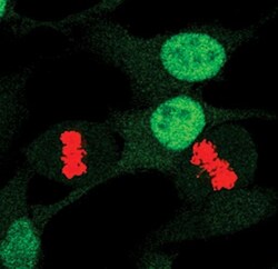
- Immunofluorescent analysis of Phospho-Cdc2 pTyr15 in asynchronous HeLa cells using a Phospho-Cdc2 pTyr15 monoclonal antibody (Product # MA5-15062) (green) and a Phospho-Histone H3 monoclonal antibody (red).

- Submitted by
- Invitrogen Antibodies (provider)
- Main image

- Experimental details
- Immunofluorescent analysis of Phospho-Cdc2 pTyr15 in asynchronous HeLa cells using a Phospho-Cdc2 pTyr15 monoclonal antibody (Product # MA5-15062) (green) and a Phospho-Histone H3 monoclonal antibody (red).

Supportive validation
- Submitted by
- Invitrogen Antibodies (provider)
- Main image
- Experimental details
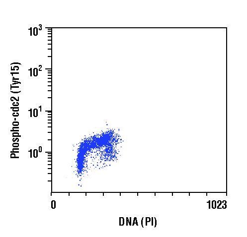
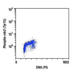
- Flow cytometric analysis of Phospho-Cdc2 pTyr15 in Jurkat cells using a Phospho-Cdc2 pTyr15 monoclonal antibody (Product # MA5-15062) versus propidium iodide (DNA content).

- Submitted by
- Invitrogen Antibodies (provider)
- Main image

- Experimental details
- Flow cytometric analysis of Phospho-Cdc2 pTyr15 in Jurkat cells using a Phospho-Cdc2 pTyr15 monoclonal antibody (Product # MA5-15062) versus propidium iodide (DNA content).