 Explore
 Validate
 Learn
LearnNBP1-32775
antibody from Novus Biologicals
Targeting: MAD1L1
HsMAD1, MAD1, PIG9, TP53I9, TXBP181
 Western blot
 Immunocytochemistry Immunoprecipitation Immunohistochemistry Chromatin Immunoprecipitation
Antibody data
- Antibody Data
- Antigen structure
- References [2]
- Comments [0]
- Validations
- Western blot [4]
- Immunoprecipitation [1]
- Immunohistochemistry [1]
Submit
Validation data
Reference
Comment
Report error
- Product number
- NBP1-32775 - Provider product page

- Provider
- Novus Biologicals
- Proper citation
- Novus Cat#NBP1-32775, RRID:AB_2139253
- Product name
- Rabbit Polyclonal MAD1L1/MAD1 Antibody
- Antibody type
- Polyclonal
- Description
- Immunogen affinity purified.
- Reactivity
- Human, Mouse, Rat
- Host
- Rabbit
- Isotype
- IgG
- Vial size
- 100 ul
- Storage
- Aliquot and store at -20C or -80C. Avoid freeze-thaw cycles.
Submitted references The spindle checkpoint protein MAD1 regulates the expression of E-cadherin and prevents cell migration.
RED, a spindle pole-associated protein, is required for kinetochore localization of MAD1, mitotic progression, and activation of the spindle assembly checkpoint.
Chen Y, Yeh PC, Huang JC, Yeh CC, Juang YL
Oncology reports 2012 Feb;27(2):487-91
Oncology reports 2012 Feb;27(2):487-91
RED, a spindle pole-associated protein, is required for kinetochore localization of MAD1, mitotic progression, and activation of the spindle assembly checkpoint.
Yeh PC, Yeh CC, Chen YC, Juang YL
The Journal of biological chemistry 2012 Apr 6;287(15):11704-16
The Journal of biological chemistry 2012 Apr 6;287(15):11704-16
No comments: Submit comment
Supportive validation
- Submitted by
- Novus Biologicals (provider)
- Main image
- Experimental details
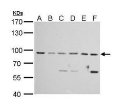
- Western Blot: MAD1L1/MAD1 Antibody [NBP1-32775] - A. 30 ug Neuro2A whole cell lysate/extract B. 30 ug GL261 whole cell lysate/extract C. 30 ug C8D30 whole cell lysate/extract D. 30 ug NIH-3T3 whole cell lysate/extract E. 30 ug BCL-1 whole cell lysate/extract F. 30 ug Raw264.7 whole cell lysate/extract 7.5% SDS-PAGE MAD1 antibody dilution: 1:3000. The HRP-conjugated anti-rabbit IgG antibody (NBP2-19301) was used to detect the primary antibody.
- Submitted by
- Novus Biologicals (provider)
- Main image
- Experimental details
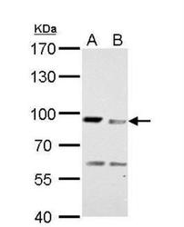
- Western Blot: MAD1L1/MAD1 Antibody [NBP1-32775] - A. 30 ug PC-12 whole cell lysate/extract B. 30 ug Rat 2 whole cell lysate/extract 7.5% SDS-PAGE MAD1 antibody dilution: 1:3000 The HRP-conjugated anti-rabbit IgG antibody (NBP2-19301) was used to detect the primary antibody.

- Submitted by
- Novus Biologicals (provider)
- Main image
- Experimental details
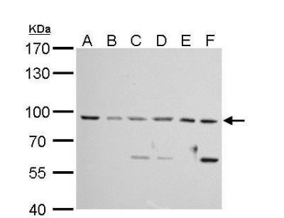
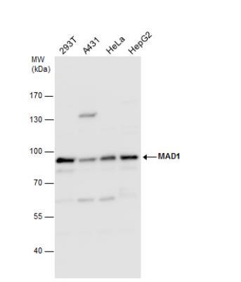
- Western Blot: MAD1L1/MAD1 Antibody [NBP1-32775] - Various whole cell extracts (30 ug) were separated by 7.5% SDS-PAGE, and the membrane was blotted with MAD1 antibody diluted by 1:5000.
- Submitted by
- Novus Biologicals (provider)
- Main image
- Experimental details
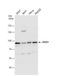
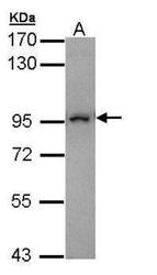
- Western Blot: MAD1L1/MAD1 Antibody [NBP1-32775] - Sample (30 ug of whole cell lysate) A: Molt-4 7.5% SDS PAGE diluted at 1:10000

Supportive validation
- Submitted by
- Novus Biologicals (provider)
- Main image
- Experimental details
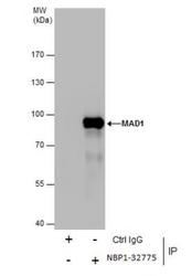
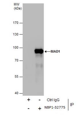
- Immunoprecipitation: MAD1L1/MAD1 Antibody [NBP1-32775] - Immunoprecipitation of MAD1 protein from HeLa whole cell extracts using 5 ug of MAD1 antibody. Western blot analysis was performed using MAD1 antibody. EasyBlot anti-Rabbit IgG was used as a secondary reagent.
Supportive validation
- Submitted by
- Novus Biologicals (provider)
- Main image
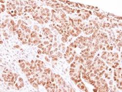
- Experimental details
- Immunohistochemistry-Paraffin: MAD1L1/MAD1 Antibody [NBP1-32775] - Paraffin-embedded SW480 Xenograft, using antibody at 1:100 dilution.
