 Explore
 Validate
 Learn
Learn Western blot
Antibody data
- Antibody Data
- Antigen structure
- References [0]
- Comments [0]
- Validations
- Western blot [3]
- Immunohistochemistry [1]
Submit
Validation data
Reference
Comment
Report error
- Product number
- PA5-27510 - Provider product page

- Provider
- Invitrogen Antibodies
- Product name
- SLC25A11 Polyclonal Antibody
- Antibody type
- Polyclonal
- Antigen
- Recombinant protein fragment
- Description
- Recommended positive controls: Jurkat, Raji, rat kidney. Predicted reactivity: Mouse (96%), Rat (96%), Zebrafish (92%), Xenopus laevis (93%), Sheep (96%), Rhesus Monkey (100%), Bovine (95%). Store product as a concentrated solution. Centrifuge briefly prior to opening the vial.
- Reactivity
- Human, Mouse, Rat
- Host
- Rabbit
- Isotype
- IgG
- Vial size
- 100 µL
- Concentration
- 1 mg/mL
- Storage
- Store at 4°C short term. For long term storage, store at -20°C, avoiding freeze/thaw cycles.
No comments: Submit comment
Supportive validation
- Submitted by
- Invitrogen Antibodies (provider)
- Main image
- Experimental details
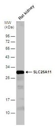
- Western blot analysis of SLC25A11 was performed by separating 50 µg of rat tissue extract by 12% SDS-PAGE. Proteins were transferred to a membrane and probed with a SLC25A11 Polyclonal Antibody (Product # PA5-27510) at a dilution of 1:1000. The HRP-conjugated anti-rabbit IgG antibody was used to detect the primary antibody.
- Submitted by
- Invitrogen Antibodies (provider)
- Main image
- Experimental details
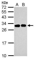
- Western Blot using SLC25A11 Polyclonal Antibody (Product # PA5-27510). Sample (30 µg of whole cell lysate). Lane A: Jurkat. Lane B: Raji. 12% SDS PAGE. SLC25A11 Polyclonal Antibody (Product # PA5-27510) diluted at 1:5,000. The HRP-conjugated anti-rabbit IgG antibody was used to detect the primary antibody.

- Submitted by
- Invitrogen Antibodies (provider)
- Main image
- Experimental details
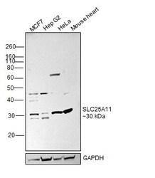
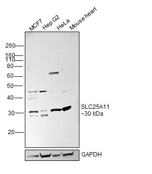
- Western blot was performed using SLC25A11 Polyclonal Antibody (Product # PA5-27510) and a 30 kDa band corresponding to SLC25A11 was observed across cell lines. Whole cell extracts (40 µg lysate) of MCF7 (Lane 1), Hep G2 (Lane 2), HeLa (Lane 3), Mouse heart (Lane 4) were electrophoresed using NuPAGE™ 12% Bis-Tris Protein Gel (Product # NP0341BOX), 10 well. Resolved proteins were then transferred onto a nitrocellulose membrane (Product # IB23001) by iBlot® 2 Dry Blotting System (Product # IB21001). The blot was probed with the primary antibody (1:1000 dilution) and detected by chemiluminescence with Goat anti-Rabbit IgG (H+L) Superclonal™ Recombinant Secondary Antibody, HRP (Product # A27036, 1:20,000 dilution) using the iBright™ FL1500 Imaging System (Product # A44115). Chemiluminescent detection was performed using SuperSignal™ West Atto Ultimate Sensitivity Substrate (Product # A38556).
Supportive validation
- Submitted by
- Invitrogen Antibodies (provider)
- Main image
- Experimental details
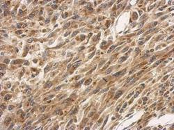
- Immunohistochemical analysis of paraffin-embedded U87 xenograft, using SLC25A11 (Product # PA5-27510) antibody at 1:500 dilution. Antigen Retrieval: EDTA based buffer, pH 8.0, 15 min.
