 Explore
 Validate
 Learn
Learn Western blot
 Immunocytochemistry
Antibody data
- Antibody Data
- Antigen structure
- References [0]
- Comments [0]
- Validations
- Western blot [2]
- Other assay [2]
Submit
Validation data
Reference
Comment
Report error
- Product number
- PA5-19561 - Provider product page

- Provider
- Invitrogen Antibodies
- Product name
- Anti-UBC9 Polyclonal Antibody
- Antibody type
- Polyclonal
- Antigen
- Synthetic peptide
- Description
- This antibody is predicted to react with chicken, Xenopus laevis, fruit fly (Drosophila melanogaster) and zebrafish based on sequence homology.
- Reactivity
- Human, Mouse, Rat
- Host
- Rabbit
- Isotype
- IgG
- Vial size
- 100 µg
- Concentration
- 0.2 mg/mL
- Storage
- Store at 4°C short term. For long term storage, store at -20°C, avoiding freeze/thaw cycles.
No comments: Submit comment
Supportive validation
- Submitted by
- Invitrogen Antibodies (provider)
- Main image
- Experimental details
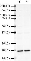
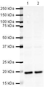
- Western blot analysis of HeLa Whole Cell Lysate using Product # PA5-19561, UBE2I/UBC9 primary antibody at a dilution of 1 µg/mL (lane 1). Staining of Jurkat Whole Cell Lysate at a dilution of 1 µg/mL (lane 2). Blot treated with a secondary IR Dye680-conjugated Goat polyclonal anti-Rabbit antibody was used at a dilution of 1:10000.
- Submitted by
- Invitrogen Antibodies (provider)
- Main image
- Experimental details
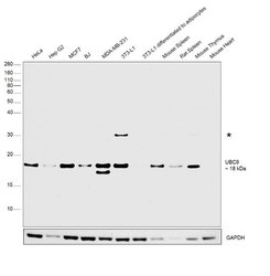
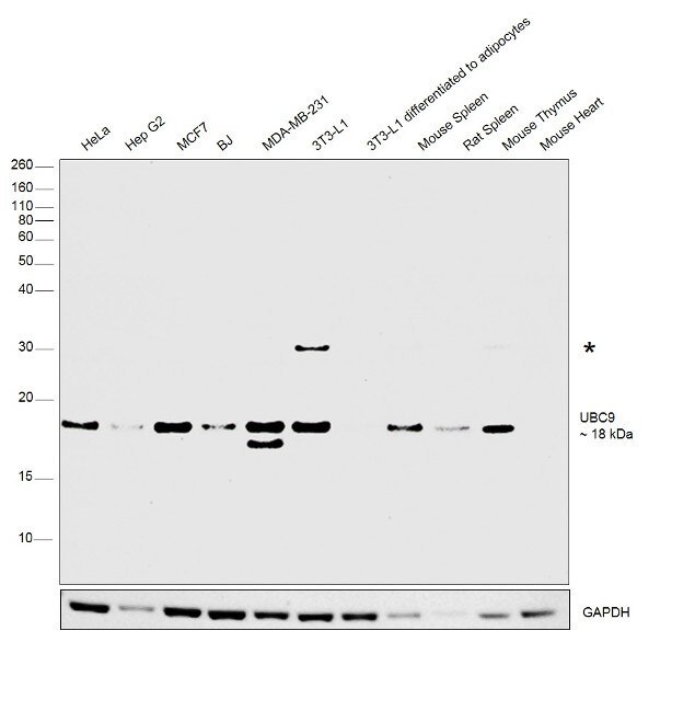
- Western blot was performed using Anti-UBC9 Polyclonal Antibody (Product # PA5-19561) and a 18 kDa band corresponding to UBC9 was observed across cell lines and tissue extracts along with an uncharacterized band (*) at ~30 kDa in the 3T3-L1 and ~17 kDa band in MDA-MB-231. Whole cell extracts (30 µg lysate) of HeLa (Lane 1), Hep-G2 (Lane 2), MCF7 (Lane 3), BJ (Lane 4), MDA-MB-231 (Lane 5), 3T3-L1 (Lane 6), 3T3-L1 differentiated to adipocytes (Lane 7), tissue extracts (30ug lysate) of Mouse Spleen (Lane 8), Rat Spleen (Lane 9), Mouse Thymus (Lane 10) and Mouse Heart (Lane 11) were electrophoresed using NuPAGE® 4-12 % Bis-Tris gel (Product # NP0322BOX). Resolved proteins were then transferred onto a nitrocellulose membrane (Product # IB23001) by iBlot® 2 Dry Blotting System (Product # IB21001). The blot was probed with the primary antibody (1:400 dilution) and detected by chemiluminescence with Goat anti-Rabbit IgG (H+L) Superclonal™ Recombinant Secondary Antibody, HRP (Product # A27036, 1:4000 dilution) using the iBright FL 1000 (Product # A32752). Chemiluminescent detection was performed using Novex® ECL Chemiluminescent Substrate Reagent Kit (Product # WP20005).
Supportive validation
- Submitted by
- Invitrogen Antibodies (provider)
- Main image
- Experimental details
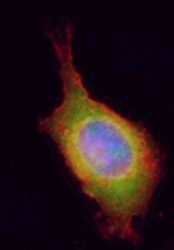
- Immunofluorescent staining of HeLa using Product # PA5-19561, anti-UBE2I/UBC9 antibody. The cells were fixed with PFA (4%) for 10 minutes, permabilised with BSA (1%), normal goat serum (10%) and glycine (0.3 M) in 0.1% T-BST for 20 minutes and exposed to the primary antibody at a concentration of 1 µg/mL for 1 hour at room temp. The secondary antibody was a 448 fluorescence conjugated Goat anti-rabbit IgG (green) at a dilution of 1:1000. A WGA- 594 fluorescent conjugated stain was used to label plasma membranes (red) and the nuclei stain was DAPI (blue).

- Submitted by
- Invitrogen Antibodies (provider)
- Main image
- Experimental details
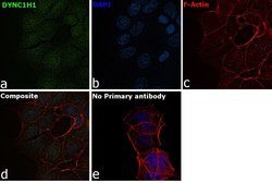
- Immunofluorescence analysis of UBC9 was performed using 70% confluent log phase MCF7 cells. The cells were fixed with 4% paraformaldehyde for 10 minutes, permeabilized with 0.1% Triton™ X-100 for 15 minutes, and blocked with 2% BSA for 1 hour at room temperature. The cells were labeled with UBC9 Polyclonal Antibody (Product # PA5-19561) at 1µg/ml in 0.1% BSA, incubated at 4 degree Celsius overnight and then labeled with Goat anti-Rabbit IgG (H+L) Superclonal™ Recombinant Secondary Antibody, Alexa Fluor® 488 conjugate (Product # A27034) at a dilution of 1:2000 for 45 minutes at room temperature (Panel a: green). Nuclei (Panel b: blue) were stained with SlowFade® Gold Antifade Mountant with DAPI (Product # S36938). F-actin (Panel c: red) was stained with Rhodamine Phalloidin (Product # R415, 1:300). Panel d represents the merged image showing localization to the nucleus and cytoplasm. Panel e represents control cells with no primary antibody to assess background. The images were captured at 60X magnification.
