Explore
 Validate
 Learn
Learn Western blot
 ELISA
ELISAAntibody data
- Antibody Data
- Antigen structure
- References [8]
- Comments [0]
- Validations
- Western blot [1]
- Immunocytochemistry [1]
- Immunohistochemistry [1]
Submit
Validation data
Reference
Comment
Report error
- Product number
- 11666-1-AP - Provider product page

- Provider
- Proteintech Group
- Proper citation
- Proteintech Cat#11666-1-AP, RRID:AB_10949076
- Product name
- WSB1 antibody
- Antibody type
- Polyclonal
- Description
- WSB1 antibody (Cat. #11666-1-AP) is a rabbit polyclonal antibody that shows reactivity with human and has been validated for the following applications: IF, IHC, WB, ELISA.
- Reactivity
- Human
- Host
- Rabbit
- Conjugate
- Unconjugated
- Isotype
- IgG
- Vial size
- 20ul, 150ul
Submitted references WSB1 promotes prostate cancer malignancy through OTUD4-mediated ISOC2 stabilization and P16INK4a suppression.
S-nitrosylation of pVHL regulates β(2) adrenergic receptor function.
circWSB1 promotes tumor progression in ccRCC via circWSB1/miR-182-5p/WSB1 axis.
Single-cell protein activity analysis reveals a novel subpopulation of chondrocytes and the corresponding key master regulator proteins associated with anti-senescence and OA progression.
BDE209-promoted Dio2 degradation in H4 glioma cells through the autophagy pathway, resulting in hypothyroidism and leading to neurotoxicity.
WSB1 overcomes oncogene-induced senescence by targeting ATM for degradation.
Hypoxia-Induced WSB1 Promotes the Metastatic Potential of Osteosarcoma Cells.
WSB1 promotes tumor metastasis by inducing pVHL degradation.
Sun J, Wang F, Feng Y, Qiu X, Qu W, He X, Xie J, Li G
American journal of cancer research 2025;15(10):4434-4451
American journal of cancer research 2025;15(10):4434-4451
S-nitrosylation of pVHL regulates β(2) adrenergic receptor function.
Grimmett ZW, Hayashi H, Raffay TM, Lin J, Premont RT, Stamler JS
Proceedings of the National Academy of Sciences of the United States of America 2025 Sep 16;122(37):e2515326122
Proceedings of the National Academy of Sciences of the United States of America 2025 Sep 16;122(37):e2515326122
circWSB1 promotes tumor progression in ccRCC via circWSB1/miR-182-5p/WSB1 axis.
Tang G, Liu J, Gao X, Tang W, Chen J, Wu M, Lv Z, Zhang Y, Cai Y, Qi L
International journal of biological macromolecules 2024 Jan;256(Pt 1):128338
International journal of biological macromolecules 2024 Jan;256(Pt 1):128338
Single-cell protein activity analysis reveals a novel subpopulation of chondrocytes and the corresponding key master regulator proteins associated with anti-senescence and OA progression.
Guang Z, Min Z, Jun-Tan L, Tian-Xu D, Xiang G
Frontiers in immunology 2023;14:1077003
Frontiers in immunology 2023;14:1077003
BDE209-promoted Dio2 degradation in H4 glioma cells through the autophagy pathway, resulting in hypothyroidism and leading to neurotoxicity.
Liu M, Yu Z, Yang F, Zhao Z, Zhou M, Wang C, Zhang B, Liang G, Liu X, Shao J
Toxicology 2023 Aug 1;494:153581
Toxicology 2023 Aug 1;494:153581
WSB1 overcomes oncogene-induced senescence by targeting ATM for degradation.
Kim JJ, Lee SB, Yi SY, Han SA, Kim SH, Lee JM, Tong SY, Yin P, Gao B, Zhang J, Lou Z
Cell research 2017 Feb;27(2):274-293
Cell research 2017 Feb;27(2):274-293
Hypoxia-Induced WSB1 Promotes the Metastatic Potential of Osteosarcoma Cells.
Cao J, Wang Y, Dong R, Lin G, Zhang N, Wang J, Lin N, Gu Y, Ding L, Ying M, He Q, Yang B
Cancer research 2015 Nov 15;75(22):4839-51
Cancer research 2015 Nov 15;75(22):4839-51
WSB1 promotes tumor metastasis by inducing pVHL degradation.
Kim JJ, Lee SB, Jang J, Yi SY, Kim SH, Han SA, Lee JM, Tong SY, Vincelette ND, Gao B, Yin P, Evans D, Choi DW, Qin B, Liu T, Zhang H, Deng M, Jen J, Zhang J, Wang L, Lou Z
Genes & development 2015 Nov 1;29(21):2244-57
Genes & development 2015 Nov 1;29(21):2244-57
No comments: Submit comment
Supportive validation
- Submitted by
- Proteintech Group (provider)
- Main image
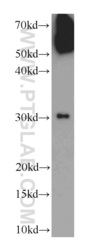
- Experimental details
- SGC-7901 cells were subjected to SDS PAGE followed by western blot with 11666-1-AP(WSB1 antibody) at dilution of 1:1000
- Sample type
- cell line

Supportive validation
- Submitted by
- Proteintech Group (provider)
- Main image
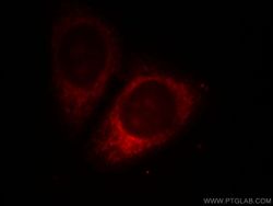
- Experimental details
- Immunofluorescent analysis of HepG2 cells, using WSB1 antibody 11666-1-AP at 1:25 dilution and Rhodamine-labeled goat anti-rabbit IgG (red).
- Sample type
- cell line

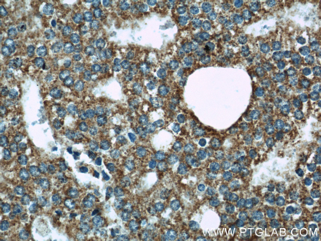
Supportive validation
- Submitted by
- Proteintech Group (provider)
- Main image
- Experimental details
- Immunohistochemical of paraffin-embedded human prostate cancer using 11666-1-AP(WSB1 antibody) at dilution of 1:50 (under 40x lens)
- Sample type
- tissue