Explore
 Validate
 Learn
Learn Western blot
Antibody data
- Antibody Data
- Antigen structure
- References [1]
- Comments [0]
- Validations
- Western blot [1]
- Immunocytochemistry [1]
- Immunohistochemistry [1]
- Flow cytometry [1]
Submit
Validation data
Reference
Comment
Report error
- Product number
- AP6709a - Provider product page

- Provider
- Abcepta
- Proper citation
- Abgent Cat#AP6709a, RRID:AB_1968354
- Product name
- TIMP1 Antibody (N-term)
- Antibody type
- Polyclonal
- Antigen
- Synthetic peptide
- Description
- Purified Rabbit Polyclonal Antibody (Pab)
- Reactivity
- Human
- Host
- Rabbit
- Isotype
- IgG
- Vial size
- 400 µl
- Concentration
- 2 mg/ml
- Storage
- Maintain refrigerated at 2-8°C for up to 6 months. For long term storage store at -20°C in small aliquots to prevent freeze-thaw cycles.
Submitted references A lipoxin A4 analog ameliorates blood-brain barrier dysfunction and reduces MMP-9 expression in a rat model of focal cerebral ischemia-reperfusion injury.
Wu Y, Wang YP, Guo P, Ye XH, Wang J, Yuan SY, Yao SL, Shang Y
Journal of molecular neuroscience : MN 2012 Mar;46(3):483-91
Journal of molecular neuroscience : MN 2012 Mar;46(3):483-91
No comments: Submit comment
Supportive validation
- Submitted by
- Abcepta (provider)
- Main image

- Experimental details
- Western blot analysis of TIMP1 Antibody (N-term) (Cat. #AP6709a) in CEM cell line lysates (35ug/lane). TIMP1 (arrow) was detected using the purified Pab.
- Primary Ab dilution
- 1:1000

Supportive validation
- Submitted by
- Abcepta (provider)
- Main image

- Experimental details
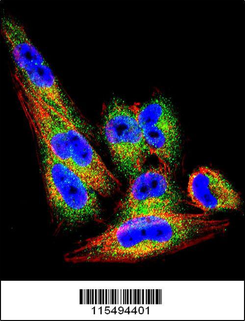
- Confocal immunofluorescent analysis of TIMP1 Antibody (N-term)(Cat#AP6709a) with A2058 cell followed by Alexa Fluor 488-conjugated goat anti-rabbit lgG (green). Actin filaments have been labeled with Alexa Fluor 555 phalloidin (red).DAPI was used to stain the cell nuclear (blue).
- Primary Ab dilution
- 1:10~50
Supportive validation
- Submitted by
- Abcepta (provider)
- Main image
- Experimental details
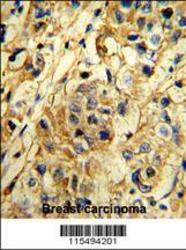
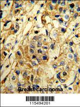
- "Formalin-fixed and paraffin-embedded human breast carcinoma reacted with TIMP1 Antibody (N-term), which was peroxidase-conjugated to the secondary antibody, followed by DAB staining. This data demonstrates the use of this antibody for immunohistochemistry; clinical relevance has not been evaluated."
- Primary Ab dilution
- 1:10~50
Supportive validation
- Submitted by
- Abcepta (provider)
- Main image
- Experimental details
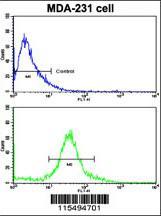
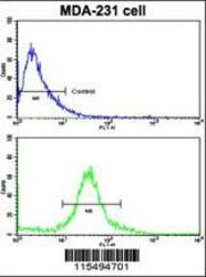
- Flow cytometric analysis of MDA-231 cells using TIMP1 Antibody (N-term)(bottom histogram) compared to a negative control cell (top histogram). FITC-conjugated goat-anti-rabbit secondary antibodies were used for the analysis.
- Primary Ab dilution
- 1:10~50