Explore
 Validate
 Learn
Learn Western blot
Antibody data
- Antibody Data
- Antigen structure
- References [7]
- Comments [0]
- Validations
- Western blot [4]
- Immunohistochemistry [1]
- Blocking/Neutralizing [1]
Submit
Validation data
Reference
Comment
Report error
- Product number
- AF970 - Provider product page

- Provider
- R&D Systems
- Product name
- Human TIMP-1 Antibody
- Antibody type
- Polyclonal
- Description
- Antigen Affinity-purified. Detects human TIMP-1 in direct ELISAs and Western blots. In direct ELISAs, approximately 20% cross-reactivity with recombinant mouse TIMP-1 and recombinant rat TIMP-1 is observed and less than 1% cross-reactivity with recombinant human (rh) TIMP-2, rhTIMP-3, and rhTIMP-4 is observed.
- Reactivity
- Human
- Host
- Goat
- Conjugate
- Unconjugated
- Antigen sequence
Q6FGX5- Isotype
- IgG
- Vial size
- 100 ug
- Concentration
- LYOPH
- Storage
- Use a manual defrost freezer and avoid repeated freeze-thaw cycles. 12 months from date of receipt, -20 to -70 °C as supplied. 1 month, 2 to 8 °C under sterile conditions after reconstitution. 6 months, -20 to -70 °C under sterile conditions after reconstitution.
Submitted references Targeting TR4 nuclear receptor suppresses prostate cancer invasion via reduction of infiltrating macrophages with alteration of the TIMP-1/MMP2/MMP9 signals.
Plasma levels of the MMP-9:TIMP-1 complex as prognostic biomarker in breast cancer: a retrospective study.
Adipocytes promote ovarian cancer metastasis and provide energy for rapid tumor growth.
Breast cancer cells induce cancer-associated fibroblasts to secrete hepatocyte growth factor to enhance breast tumorigenesis.
Primary human acute myelogenous leukemia cells release matrix metalloproteases and their inhibitors: release profile and pharmacological modulation.
Fibroblast-conditioned media promote human sarcoma cell invasion.
Coregulation of vascular tube stabilization by endothelial cell TIMP-2 and pericyte TIMP-3.
Ding X, Yang DR, Xia L, Chen B, Yu S, Niu Y, Wang M, Li G, Chang C
Molecular cancer 2015 Jan 27;14(1):16
Molecular cancer 2015 Jan 27;14(1):16
Plasma levels of the MMP-9:TIMP-1 complex as prognostic biomarker in breast cancer: a retrospective study.
Thorsen SB, Christensen SL, Würtz SO, Lundberg M, Nielsen BS, Vinther L, Knowles M, Gee N, Fredriksson S, Møller S, Brünner N, Schrohl AS, Stenvang J
BMC cancer 2013 Dec 13;13:598
BMC cancer 2013 Dec 13;13:598
Adipocytes promote ovarian cancer metastasis and provide energy for rapid tumor growth.
Nieman KM, Kenny HA, Penicka CV, Ladanyi A, Buell-Gutbrod R, Zillhardt MR, Romero IL, Carey MS, Mills GB, Hotamisligil GS, Yamada SD, Peter ME, Gwin K, Lengyel E
Nature medicine 2011 Oct 30;17(11):1498-503
Nature medicine 2011 Oct 30;17(11):1498-503
Breast cancer cells induce cancer-associated fibroblasts to secrete hepatocyte growth factor to enhance breast tumorigenesis.
Tyan SW, Kuo WH, Huang CK, Pan CC, Shew JY, Chang KJ, Lee EY, Lee WH
PloS one 2011 Jan 13;6(1):e15313
PloS one 2011 Jan 13;6(1):e15313
Primary human acute myelogenous leukemia cells release matrix metalloproteases and their inhibitors: release profile and pharmacological modulation.
Reikvam H, Hatfield KJ, Oyan AM, Kalland KH, Kittang AO, Bruserud O
European journal of haematology 2010 Mar;84(3):239-51
European journal of haematology 2010 Mar;84(3):239-51
Fibroblast-conditioned media promote human sarcoma cell invasion.
Bittner JG 4th, Wilson M, Shah MB, Albo D, Feig BW, Wang TN
Surgery 2009 Jan;145(1):42-7
Surgery 2009 Jan;145(1):42-7
Coregulation of vascular tube stabilization by endothelial cell TIMP-2 and pericyte TIMP-3.
Saunders WB, Bohnsack BL, Faske JB, Anthis NJ, Bayless KJ, Hirschi KK, Davis GE
The Journal of cell biology 2006 Oct 9;175(1):179-91
The Journal of cell biology 2006 Oct 9;175(1):179-91
No comments: Submit comment
Supportive validation
- Submitted by
- R&D Systems (provider)
- Main image
- Experimental details
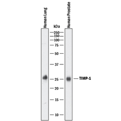
- Detection of Human TIMP-1 by Western Blot. Western blot shows lysates of human lung tissue and human prostate tissue. PVDF membrane was probed with 1 µg/mL of Goat Anti-Human TIMP-1 Antigen Affinity-purified Polyclonal Antibody (Catalog # AF970) followed by HRP-conjugated Anti-Goat IgG Secondary Antibody (Catalog # HAF017). A specific band was detected for TIMP-1 at approximately 25 kDa (as indicated). This experiment was conducted under reducing conditions and using Immunoblot Buffer Group 1.

- Submitted by
- R&D Systems (provider)
- Main image
- Experimental details
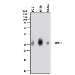
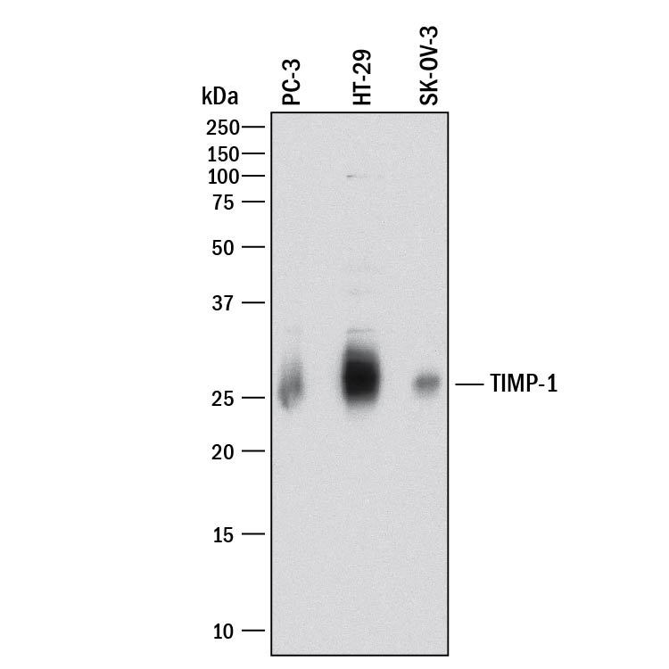
- Detection of Human TIMP-1 by Western Blot. Western blot shows lysates of PC-3 human prostate cancer cell line, HT-29 human colon adenocarcinoma cell line, and SK-OV-3 human ovarian adenocarcinoma cell line. PVDF membrane was probed with 5 µg/mL of Goat Anti-Human TIMP-1 Antigen Affinity-purified Polyclonal Antibody (Catalog # AF970) followed by HRP-conjugated Anti-Goat IgG Secondary Antibody (Catalog # HAF017). A specific band was detected for TIMP-1 at approximately 26 kDa (as indicated). This experiment was conducted under reducing conditions and using Immunoblot Buffer Group 1.
- Submitted by
- R&D Systems (provider)
- Main image
- Experimental details
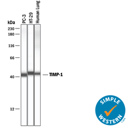
- Detection of Human TIMP-1 by Simple WesternTM. Simple Western lane view shows lysates of PC-3 human prostate cancer cell line, HT-29 human colon adenocarcinoma cell line, and human lung tissue, loaded at 0.2 mg/mL. A specific band was detected for TIMP-1 at approximately 42-45 kDa (as indicated) using 50 µg/mL of Goat Anti-Human TIMP-1 Antigen Affinity-purified Polyclonal Antibody (Catalog # AF970) followed by 1:50 dilution of HRP-conjugated Anti-Goat IgG Secondary Antibody (Catalog # HAF109). This experiment was conducted under reducing conditions and using the 12-230 kDa separation system.

- Submitted by
- R&D Systems (provider)
- Main image
- Experimental details
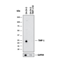
- Western Blot Shows Human TIMP-1 Specificity by Using Knockout Cell Line. Western blot shows lysates of SK-OV-3 human ovarian adenocarcinoma cell line and TIMP-1 knockout SK-OV-3 cell line (KO). PVDF membrane was probed with 2 µg/mL of Goat Anti-Human TIMP-1 Antigen Affinity-purified Polyclonal Antibody (Catalog # AF970) followed by HRP-conjugated Anti-Goat IgG Secondary Antibody (Catalog # HAF017). A specific band was detected for TIMP-1 at approximately 25 kDa (as indicated) in the parental SK-OV-3 cell line, but is not detectable in knockout SK-OV-3 cell line. GAPDH (Catalog # AF5718) is shown as a loading control. This experiment was conducted under reducing conditions and using Immunoblot Buffer Group 1.

Supportive validation
- Submitted by
- R&D Systems (provider)
- Main image
- Experimental details
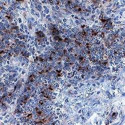
- TIMP-1 in Human Colon Cancer Tissue. TIMP-1 was detected in immersion fixed paraffin-embedded sections of human colon cancer tissue using Goat Anti-Human TIMP-1 Antigen Affinity-purified Polyclonal Antibody (Catalog # AF970) at 1 µg/mL for 1 hour at room temperature followed by incubation with the Anti-Goat IgG VisUCyte™ HRP Polymer Antibody (Catalog # VC004). Before incubation with the primary antibody, tissue was subjected to heat-induced epitope retrieval using Antigen Retrieval Reagent-Basic (Catalog # CTS013). Tissue was stained using DAB (brown) and counterstained with hematoxylin (blue). Specific staining was localized to cytoplasm and extracellular space. View our protocol for IHC Staining with VisUCyte HRP Polymer Detection Reagents.

Supportive validation
- Submitted by
- R&D Systems (provider)
- Main image
- Experimental details
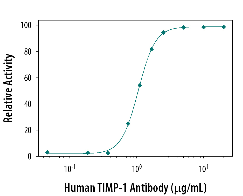
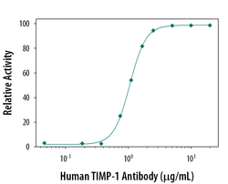
- Neutralization of TIMP-1 Activity by Human TIMP-1 Antibody. Recombinant Human MMP-2 (0.2 µg/mL, Catalog # 902-MP) activity is measured in the presence of Recombinant Human TIMP-1 (0.1 µg/mL, Catalog # 970-TM) that has been preincubated with increasing concentrations of Goat Anti-Human TIMP-1 Antigen Affinity-purified Polyclonal Antibody (Catalog # AF970). The ND50 is typically 1 µg/mL.