Explore
 Validate
 Learn
Learn Western blot
 Immunocytochemistry
Antibody data
- Antibody Data
- Antigen structure
- References [0]
- Comments [0]
- Validations
- Immunocytochemistry [3]
- Immunohistochemistry [3]
- Other assay [1]
Submit
Validation data
Reference
Comment
Report error
- Product number
- PA5-31164 - Provider product page

- Provider
- Invitrogen Antibodies
- Product name
- TUB Polyclonal Antibody
- Antibody type
- Polyclonal
- Antigen
- Recombinant full-length protein
- Description
- Recommended positive controls: NT2D1, PC-3, U87-MG, SK-N-SH, Neuro2A, GL261. Predicted reactivity: Mouse (92%), Rat (93%), Bovine (89%). Store product as a concentrated solution. Centrifuge briefly prior to opening the vial.
- Reactivity
- Human, Mouse, Rat
- Host
- Rabbit
- Isotype
- IgG
- Vial size
- 100 μL
- Concentration
- 1 mg/mL
- Storage
- Store at 4°C short term. For long term storage, store at -20°C, avoiding freeze/thaw cycles.
No comments: Submit comment
Supportive validation
- Submitted by
- Invitrogen Antibodies (provider)
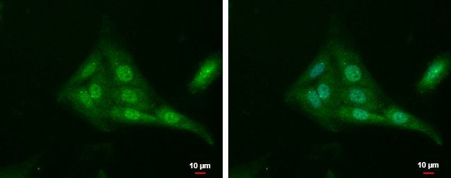
- Main image
- Experimental details
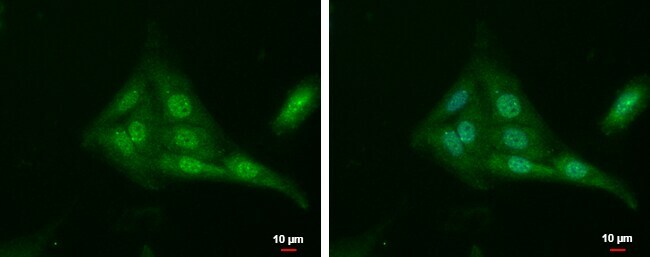
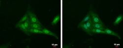
- TUB Polyclonal Antibody detects Tub protein at cytoplasm and nucleus by immunofluorescent analysis. Sample: SKNSH cells were fixed in 4% paraformaldehyde at RT for 15 min. Green: Tub protein stained by TUB Polyclonal Antibody (Product # PA5-31164) diluted at 1:500. Blue: Hoechst 33342 staining. Scale bar = 10 µm.
- Submitted by
- Invitrogen Antibodies (provider)
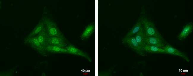
- Main image
- Experimental details
- TUB Polyclonal Antibody detects Tub protein at cytoplasm and nucleus by immunofluorescent analysis. Sample: SKNSH cells were fixed in 4% paraformaldehyde at RT for 15 min. Green: Tub protein stained by TUB Polyclonal Antibody (Product # PA5-31164) diluted at 1:500. Blue: Hoechst 33342 staining. Scale bar = 10 µm.

- Submitted by
- Invitrogen Antibodies (provider)
- Main image

- Experimental details
- TUB Polyclonal Antibody detects Tub protein at cytoplasm and nucleus by immunofluorescent analysis. Sample: SKNSH cells were fixed in 4% paraformaldehyde at RT for 15 min. Green: Tub protein stained by TUB Polyclonal Antibody (Product # PA5-31164) diluted at 1:500. Blue: Hoechst 33342 staining. Scale bar = 10 µm.
Supportive validation
- Submitted by
- Invitrogen Antibodies (provider)
- Main image
- Experimental details
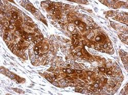
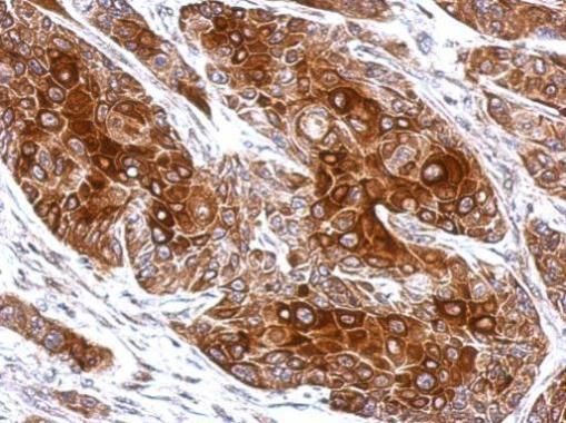
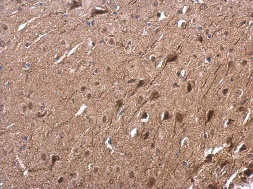
- Immunohistochemical analysis of paraffin-embedded Cal27 xenograft, using Tub (Product # PA5-31164) antibody at 1:500 dilution. Antigen Retrieval: EDTA based buffer, pH 8.0, 15 min.
- Submitted by
- Invitrogen Antibodies (provider)
- Main image
- Experimental details
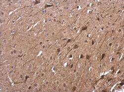
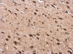
- TUB Polyclonal Antibody detects Tub protein at nucleus on mouse fore brain by immunohistochemical analysis. Sample: Paraffin-embedded mouse fore brain. TUB Polyclonal Antibody (Product # PA5-31164) dilution: 1:500. Antigen Retrieval: EDTA based buffer, pH 8.0, 15 min.
- Submitted by
- Invitrogen Antibodies (provider)
- Main image
- Experimental details
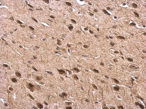
- TUB Polyclonal Antibody detects Tub protein at nucleus on rat fore brain by immunohistochemical analysis. Sample: Paraffin-embedded rat fore brain. TUB Polyclonal Antibody (Product # PA5-31164) dilution: 1:500. Antigen Retrieval: EDTA based buffer, pH 8.0, 15 min.
Supportive validation
- Submitted by
- Invitrogen Antibodies (provider)
- Main image
- Experimental details
- Immunofluorescent analysis of Tub showing staining in the cytoplasm and nucleus of SKNSH cells. SKNSH cells were fixed in 4% paraformaldehyde at RT for 15 min and stained using a Tub polyclonal antibody (Product # PA5-31164) diluted at 1:500. Blue: Hoechst 33342 staining. Scale bar = 10µm.