Explore
 Validate
 Learn
Learn Western blot
 Immunocytochemistry
Antibody data
- Antibody Data
- Antigen structure
- References [1]
- Comments [0]
- Validations
- Western blot [1]
- Immunohistochemistry [2]
Submit
Validation data
Reference
Comment
Report error
- Product number
- AF958 - Provider product page

- Provider
- Novus Biologicals
- Product name
- Goat Polyclonal GDF-3 Antibody
- Antibody type
- Polyclonal
- Description
- Immunogen affinity purified. Detects human and mouse GDF-3 in direct ELISAs and Western blots. In Western blots, less than 5% cross-reactivity with recombinant mouse (rm) GDF-5, rmGDF-6, rmGDF-7, rmGDF-8, and rmGDF-9 is observed.
- Reactivity
- Human, Mouse
- Host
- Goat
- Isotype
- IgG
- Vial size
- 100 ug
- Concentration
- LYOPH
- Storage
- Use a manual defrost freezer and avoid repeated freeze-thaw cycles. 12 months from date of receipt, -20 to -70 degreesC as supplied. 1 month, 2 to 8 degreesC under sterile conditions after reconstitution. 6 months, -20 to -70 degreesC under sterile conditions after reconstitution.
Submitted references GDF3 is a BMP inhibitor that can activate Nodal signaling only at very high doses.
Levine AJ, Levine ZJ, Brivanlou AH
Developmental biology 2009 Jan 1;325(1):43-8
Developmental biology 2009 Jan 1;325(1):43-8
No comments: Submit comment
Supportive validation
- Submitted by
- Novus Biologicals (provider)
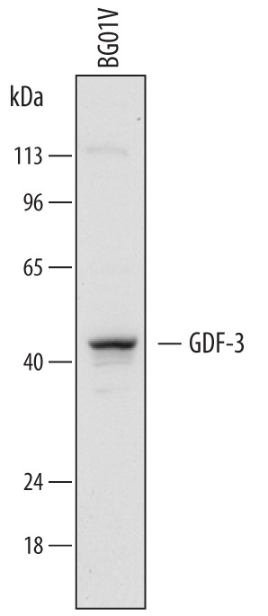
- Main image
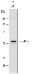
- Experimental details
- Detection of Human GDF-3 by Western Blot. Western blot shows lysates of BG01V human embryonic stem cells. PVDF membrane was probed with 1.5 µg/mL of Goat Anti-Human/Mouse GDF-3 Antigen Affinity-purified Polyclonal Antibody (Catalog # AF958) followed by HRP-conjugated Anti-Goat IgG Secondary Antibody (Catalog # HAF017). A specific band was detected for GDF-3 at approximately 42 kDa (as indicated). This experiment was conducted under reducing conditions and using Immunoblot Buffer Group 1.
Supportive validation
- Submitted by
- Novus Biologicals (provider)
- Main image
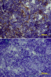
- Experimental details
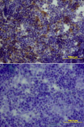
- GDF-3 in Mouse Thymus. GDF-3 was detected in perfusion fixed frozen sections of mouse thymus using Mouse GDF-3 Antigen Affinity-purified Polyclonal Antibody (Catalog # AF958) at 15 µg/mL overnight at 4 °C. Tissue was stained using the Anti-Goat HRP-DAB Cell & Tissue Staining Kit (brown; Catalog # CTS008) and counterstained with hematoxylin (blue). Lower panel shows a lack of labeling if primary antibodies are omitted and tissue is stained only with secondary antibody followed by incubation with detection reagents. View our protocol for Chromogenic IHC Staining of Frozen Tissue Sections.
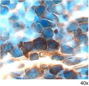
- Submitted by
- Novus Biologicals (provider)
- Main image

- Experimental details
- GDF-3 in Mouse Thymus. GDF-3 was detected in perfusion fixed frozen sections of mouse thymus using 5 µg/mL Mouse GDF-3 Antigen Affinity-purified Polyclonal Antibody (Catalog # AF958) overnight at 4 °C. Tissue was stained with the Anti-Goat HRP-DAB Cell & Tissue Staining Kit (brown; Catalog # CTS008) and counterstained with hematoxylin (blue). View our protocol for Chromogenic IHC Staining of Frozen Tissue Sections.