Explore
 Validate
 Learn
Learn Western blot
 Immunocytochemistry
Antibody data
- Antibody Data
- Antigen structure
- References [0]
- Comments [0]
- Validations
- Immunocytochemistry [2]
- Immunohistochemistry [2]
Submit
Validation data
Reference
Comment
Report error
- Product number
- PA5-47974 - Provider product page

- Provider
- Invitrogen Antibodies
- Product name
- GDF3 Polyclonal Antibody
- Antibody type
- Polyclonal
- Antigen
- Recombinant full-length protein
- Description
- In Western blots, less than 5% cross-reactivity with recombinant mouse (rm) GDF-5, rmGDF-6, rmGDF-7, rmGDF-8, and rmGDF-9 is observed. Reconstitute at 0.2 mg/mL in sterile PBS.
- Reactivity
- Human, Mouse
- Host
- Goat
- Isotype
- IgG
- Vial size
- 100 μg
- Concentration
- 0.2 mg/mL
- Storage
- -20°C, Avoid Freeze/Thaw Cycles
No comments: Submit comment
Supportive validation
- Submitted by
- Invitrogen Antibodies (provider)
- Main image
- Experimental details
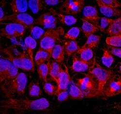
- Immunocytochemistry analysis of GDF3 in immersion fixed BG01V human embryonic stem cells. Samples were incubated in GDF3 polyclonal antibody (Product # PA5-47974) using a dilution of 10 µg/mL for 3 hours at room temperature followed by NorthernLights™ 557-conjugated Anti-Goat IgG Secondary Antibody (red) and counterstained with DAPI (blue). Specific staining was localized to cytoplasm.

- Submitted by
- Invitrogen Antibodies (provider)
- Main image

- Experimental details
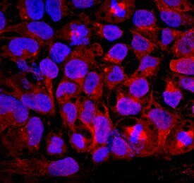
- Immunocytochemistry analysis of GDF3 in immersion fixed BG01V human embryonic stem cells. Samples were incubated in GDF3 polyclonal antibody (Product # PA5-47974) using a dilution of 10 µg/mL for 3 hours at room temperature followed by NorthernLights™ 557-conjugated Anti-Goat IgG Secondary Antibody (red) and counterstained with DAPI (blue). Specific staining was localized to cytoplasm.
Supportive validation
- Submitted by
- Invitrogen Antibodies (provider)
- Main image

- Experimental details
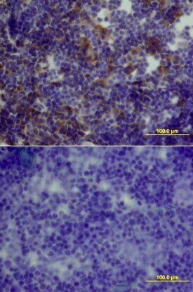
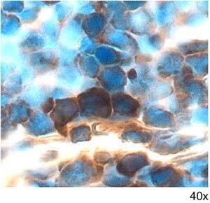
- Immunohistochemical analysis of GDF3 in perfusion fixed frozen sections of mouse thymus. Samples were incubated in GDF3 polyclonal antibody (Product # PA5-47974) using a dilution of 5 µg/mL overnight at 4 °C. Tissue was stained with the Anti-Goat HRP-DAB Cell & Tissue Staining Kit (brown) and counterstained with hematoxylin (blue).
- Submitted by
- Invitrogen Antibodies (provider)
- Main image

- Experimental details
- Immunohistochemical analysis of GDF3 in perfusion fixed frozen sections of mouse thymus. Samples were incubated in GDF3 polyclonal antibody (Product # PA5-47974) using a dilution of 15 µg/mL overnight at 4 °C. Tissue was stained using the Anti-Goat HRP-DAB Cell & Tissue Staining Kit (brown) and counterstained with hematoxylin (blue). Lower panel shows a lack of labeling if primary antibodies are omitted and tissue is stained only with secondary antibody followed by incubation with detection reagents.