 Explore
 Validate
 Learn
LearnMA5-25193
antibody from Invitrogen Antibodies
Targeting: SH3GL1
CNSA1, EEN, MGC111371, SH3D2B, SH3P8
 Western blot
 Flow cytometry
Antibody data
- Antibody Data
- Antigen structure
- References [0]
- Comments [0]
- Validations
- Western blot [1]
- Immunocytochemistry [3]
- Immunohistochemistry [1]
Submit
Validation data
Reference
Comment
Report error
- Product number
- MA5-25193 - Provider product page

- Provider
- Invitrogen Antibodies
- Product name
- SH3GL1 Monoclonal Antibody (OTI2F5)
- Antibody type
- Monoclonal
- Antigen
- Recombinant full-length protein
- Reactivity
- Human
- Host
- Mouse
- Isotype
- IgG
- Antibody clone number
- OTI2F5
- Vial size
- 100 µL
- Concentration
- 0.64 mg/mL
- Storage
- -20° C, Avoid Freeze/Thaw Cycles
No comments: Submit comment
Supportive validation
- Submitted by
- Invitrogen Antibodies (provider)
- Main image

- Experimental details
- Western blot analysis of SH3GL1 in HepG2, HeLa, HT29, A549, COS7, Jurkat, MDCK, PC12, MCF7 cells using 35 µg per lane. Samples were probed with SH3GL1 (Product # MA5-25193) monoclonal antibody.

Supportive validation
- Submitted by
- Invitrogen Antibodies (provider)
- Main image

- Experimental details
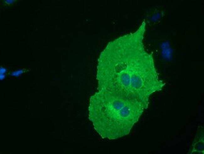
- Immunofluorescent analysis of SH3GL1 in COS7 cells. Cells were transfected with a plasmid overexpressing SH3GL1 and probed with a SH3GL1 monoclonal antibody (Product # MA5-25193).
- Submitted by
- Invitrogen Antibodies (provider)
- Main image

- Experimental details
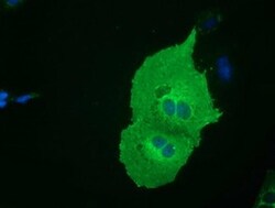
- Immunofluorescent analysis of SH3GL1 in COS7 cells. Cells were transfected with a plasmid overexpressing SH3GL1 and probed with a SH3GL1 monoclonal antibody (Product # MA5-25193).

- Submitted by
- Invitrogen Antibodies (provider)
- Main image
- Experimental details
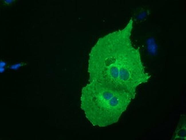
- Immunofluorescent analysis of SH3GL1 in COS7 cells. Cells were transfected with a plasmid overexpressing SH3GL1 and probed with a SH3GL1 monoclonal antibody (Product # MA5-25193).
Supportive validation
- Submitted by
- Invitrogen Antibodies (provider)
- Main image
- Experimental details
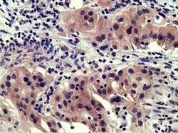
- Immunohistochemistry was performed on paraffin-embedded carcinoma of human bladder tissue. To expose target proteins, 10mM citric buffer, pH6.0, 100°C for 10min was used. Following antigen retrieval, tissues were probed with a SH3GL1 monoclonal antibody (Product # MA5-25193).
