Explore
 Validate
 Learn
Learn Western blot
Antibody data
- Antibody Data
- Antigen structure
- References [2]
- Comments [0]
- Validations
- Western blot [5]
- Immunocytochemistry [6]
- Immunohistochemistry [1]
- Chromatin Immunoprecipitation [2]
- Other assay [2]
Submit
Validation data
Reference
Comment
Report error
- Product number
- PA5-29275 - Provider product page

- Provider
- Invitrogen Antibodies
- Product name
- PPM1A Polyclonal Antibody
- Antibody type
- Polyclonal
- Antigen
- Recombinant full-length protein
- Description
- Recommended positive controls: 293T, A431, mouse brain, PC-12, Rat2. Predicted reactivity: Mouse (99%), Rat (99%), Xenopus laevis (89%), Rabbit (99%), Bovine (99%). Store product as a concentrated solution. Centrifuge briefly prior to opening the vial.
- Reactivity
- Human, Mouse, Rat
- Host
- Rabbit
- Isotype
- IgG
- Vial size
- 100 μL
- Concentration
- 1 mg/mL
- Storage
- Store at 4°C short term. For long term storage, store at -20°C, avoiding freeze/thaw cycles.
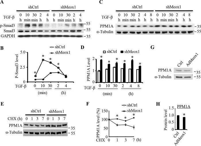
Submitted references LncRNA GAS5 attenuates fibroblast activation through inhibiting Smad3 signaling.
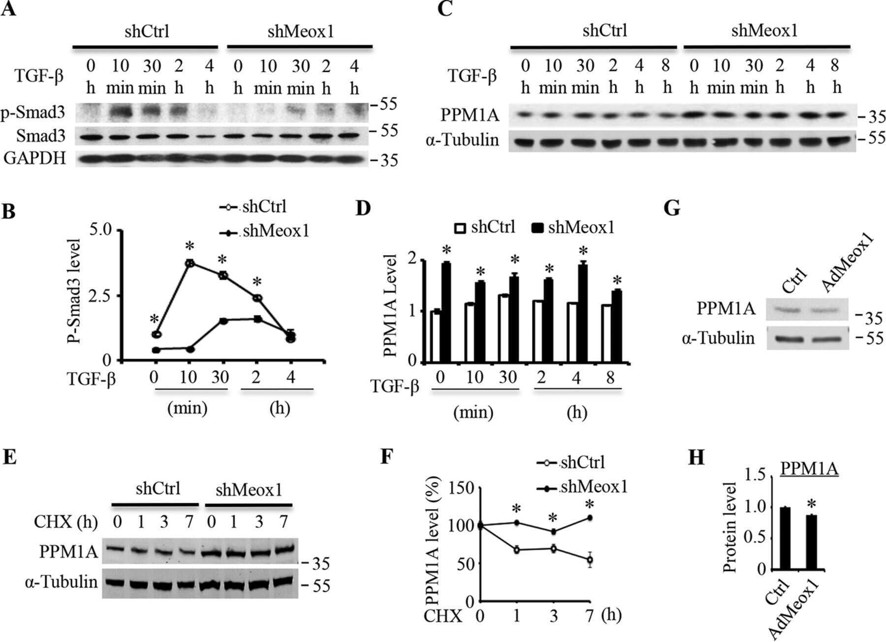
Mesenchyme homeobox 1 mediates transforming growth factor-β (TGF-β)-induced smooth muscle cell differentiation from mouse mesenchymal progenitors.
Tang R, Wang YC, Mei X, Shi N, Sun C, Ran R, Zhang G, Li W, Staveley-O'Carroll KF, Li G, Chen SY
American journal of physiology. Cell physiology 2020 Jul 1;319(1):C105-C115
American journal of physiology. Cell physiology 2020 Jul 1;319(1):C105-C115
Mesenchyme homeobox 1 mediates transforming growth factor-β (TGF-β)-induced smooth muscle cell differentiation from mouse mesenchymal progenitors.
Dong K, Guo X, Chen W, Hsu AC, Shao Q, Chen JF, Chen SY
The Journal of biological chemistry 2018 Jun 1;293(22):8712-8719
The Journal of biological chemistry 2018 Jun 1;293(22):8712-8719
No comments: Submit comment
Supportive validation
- Submitted by
- Invitrogen Antibodies (provider)
- Main image
- Experimental details
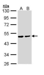
- Western Blot using PPM1A Polyclonal Antibody (Product # PA5-29275). Sample (30 µg of whole cell lysate). Lane A: 293T. Lane B: A431. 10% SDS PAGE. PPM1A Polyclonal Antibody (Product # PA5-29275) diluted at 1:1,000.

- Submitted by
- Invitrogen Antibodies (provider)
- Main image
- Experimental details
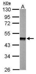
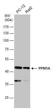
- Western Blot using PPM1A Polyclonal Antibody (Product # PA5-29275). Sample (50 µg of whole cell lysate). Lane A: Mouse brain. 10% SDS PAGE. PPM1A Polyclonal Antibody (Product # PA5-29275) diluted at 1:1,000.

- Submitted by
- Invitrogen Antibodies (provider)
- Main image
- Experimental details
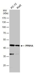
- Western Blot analysis of PPM1A was performed by separating 30 µg of various whole cell extracts by 10% SDS-PAGE. Proteins were transferred to a membrane and probed with a PPM1A Polyclonal Antibody (Product # PA5-29275) at a dilution of 1:500.
- Submitted by
- Invitrogen Antibodies (provider)
- Main image
- Experimental details
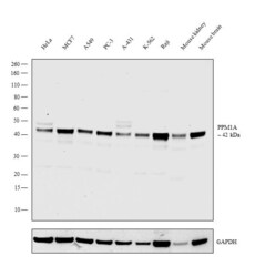
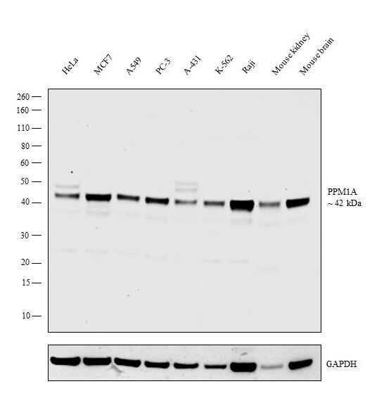
- Western blot analysis was performed on whole cell extracts (30 µg lysate) of HeLa (Lane 1), MCF7 (Lane 2), A549 (Lane 3), PC-3 (Lane 4), A-431 (Lane 5), K-562 (Lane 6), Raji (Lane 7), Mouse kidney (Lane 8), and Mouse brain (Lane 9). The blot was probed with Anti-PPM1A Polyclonal Antibody (Product # PA5-29275, 1:2000 dilution) and detected by chemiluminescence using Goat anti-Rabbit IgG (Heavy Chain) Superclonal™ Secondary Antibody, HRP conjugate (Product # A27036, 0.25 µg/mL, 1:4000 dilution). A 42 kDa band corresponding to PPM1A was observed in all cell lines tested.
- Submitted by
- Invitrogen Antibodies (provider)
- Main image
- Experimental details
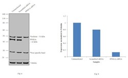
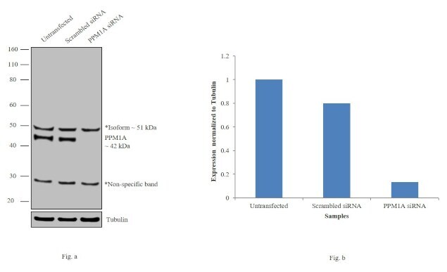
- Knockdown of PPM1A was achieved by transfecting HeLa cells with PPM1A specific siRNAs (Silencer® select Product # s10919, s10920). Western blot analysis (Fig. a) was performed using whole cell extracts from the PPM1A knockdown cells (lane 3), non-specific scrambled siRNA transfected cells (lane 2) and untransfected cells (lane 1). The blots were probed with PPM1A Polyclonal Antibody (Product # PA5-29275, 1:1000 dilution) and Goat anti-Rabbit IgG (Heavy Chain) Superclonal™ Secondary Antibody, HRP conjugate (Product # A27036, 0.25 µg/mL, 1:4000 dilution). Densitometric analysis of this western blot is shown in histogram (Fig. b). Decrease in signal upon siRNA mediated knock down confirms that antibody is specific to PPM1A. A 51KDa band corresponding to its isoform was observed along with an uncharacterized band at ~29KDa.
Supportive validation
- Submitted by
- Invitrogen Antibodies (provider)
- Main image

- Experimental details
- Immunofluorescent analysis of PPM1A showing staining in the cytoplasm of A431 cells. A431 cells were fixed in ice-cold MeOH for 5 min and stained using a PPM1A polyclonal antibody (Product # PA5-29275) diluted at 1:500. Blue: Hoechst 33342 staining.

- Submitted by
- Invitrogen Antibodies (provider)
- Main image

- Experimental details
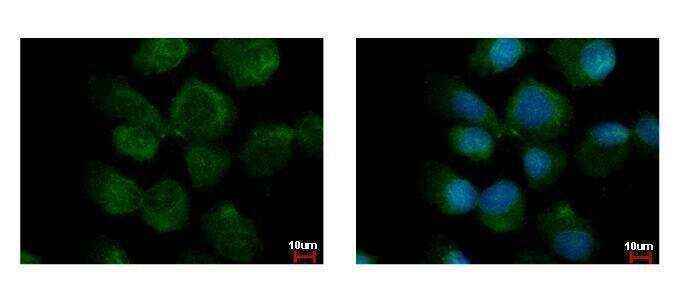
- PPM1A Polyclonal Antibody detects PPM1A protein at cytoplasm by immunofluorescent analysis. Sample: A431 cells were fixed in ice-cold MeOH for 5 min. Green: PPM1A protein stained by PPM1A Polyclonal Antibody (Product # PA5-29275) diluted at 1:500. Blue: Hoechst 33342 staining.
- Submitted by
- Invitrogen Antibodies (provider)
- Main image
- Experimental details
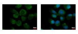
- PPM1A Polyclonal Antibody detects PPM1A protein at cytoplasm by immunofluorescent analysis. Sample: A431 cells were fixed in ice-cold MeOH for 5 min. Green: PPM1A protein stained by PPM1A Polyclonal Antibody (Product # PA5-29275) diluted at 1:500. Blue: Hoechst 33342 staining.
- Submitted by
- Invitrogen Antibodies (provider)
- Main image
- Experimental details
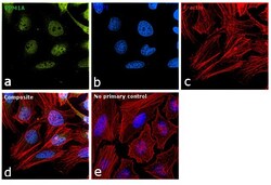
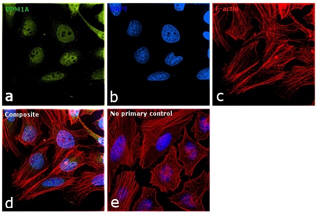
- Immunofluorescence analysis of PPM1A was performed using 70% confluent log phase HeLa cells. The cells were fixed with 4% paraformaldehyde for 10 minutes, permeabilized with 0.1% Triton™ X-100 for 15 minutes, and blocked with 1% BSA for 1 hour at room temperature. The cells were labeled with PPM1A Polyclonal Antibody (Product # PA5-29275) at 1:200 dilution in 0.1% BSA, incubated at 4 degree Celsius overnight and then labeled with Goat anti-Rabbit IgG (H+L) Superclonal™ Secondary Antibody, Alexa Fluor® 488 conjugate (Product # A27034) for 45 minutes at room temperature (Panel a: green). Nuclei (Panel b: blue) were stained with ProLong™ Diamond Antifade Mountant with DAPI (Product # P36962). F-actin (Panel c: red) was stained with Rhodamine Phalloidin (Product # R415, 1:300). Panel d represents the merged image showing predominant nuclear localization along with cytoplasm. Panel e represents control cells with no primary antibody to assess background. The images were captured at 60X magnification.

- Submitted by
- Invitrogen Antibodies (provider)
- Main image

- Experimental details
- Immunofluorescence analysis of PPM1A was performed using 70% confluent log phase HeLa cells. The cells were fixed with 4% paraformaldehyde for 10 minutes, permeabilized with 0.1% Triton™ X-100 for 15 minutes, and blocked with 1% BSA for 1 hour at room temperature. The cells were labeled with PPM1A Polyclonal Antibody (Product # PA5-29275) at 1:200 dilution in 0.1% BSA, incubated at 4 degree Celsius overnight and then labeled with Goat anti-Rabbit IgG (Heavy Chain) Superclonal™ Secondary Antibody, Alexa Fluor® 488 conjugate (Product # A27034) for 45 minutes at room temperature (Panel a: green). Nuclei (Panel b: blue) were stained with ProLong™ Diamond Antifade Mountant with DAPI (Product # P36962). F-actin (Panel c: red) was stained with Rhodamine Phalloidin (Product # R415, 1:300). Panel d represents the merged image showing predominant nuclear localization along with cytoplasm. Panel e represents control cells with no primary antibody to assess background. The images were captured at 60X magnification.
- Submitted by
- Invitrogen Antibodies (provider)
- Main image

- Experimental details
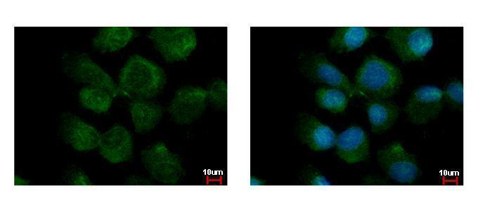
- PPM1A Polyclonal Antibody detects PPM1A protein at cytoplasm by immunofluorescent analysis. Sample: A431 cells were fixed in ice-cold MeOH for 5 min. Green: PPM1A protein stained by PPM1A Polyclonal Antibody (Product # PA5-29275) diluted at 1:500. Blue: Hoechst 33342 staining.

Supportive validation
- Submitted by
- Invitrogen Antibodies (provider)
- Main image
- Experimental details
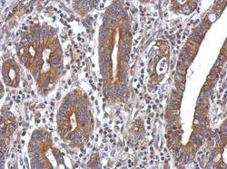
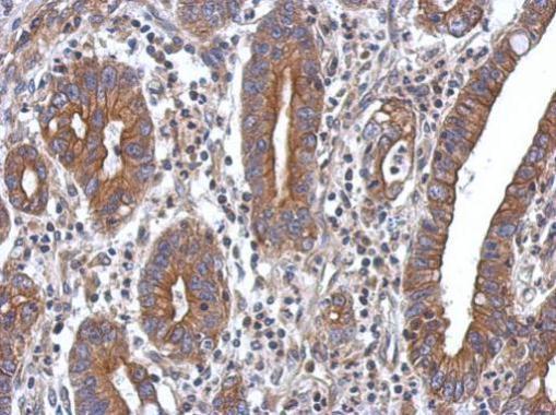
- Immunohistochemical analysis of paraffin-embedded human colon carcinoma, using PPM1A (Product # PA5-29275) antibody at 1:500 dilution. Antigen Retrieval: EDTA based buffer, pH 8.0, 15 min.
Supportive validation
- Submitted by
- Invitrogen Antibodies (provider)
- Main image

- Experimental details
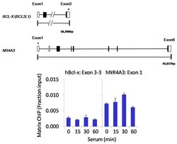
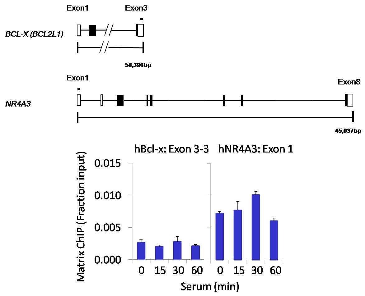
- Chromatin immunoprecipitation analysis of PPM1A was performed using cross-linked chromatin from 1x10^6 HCT116 colon carcinoma cells treated with serum for 0, 15, 30, and 60 minutes. Immunoprecipitation was performed using a multiplex microplate Matrix ChIP assay (see reference for Matrix ChIP protocol: http://www.ncbi.nlm.nih.gov/pubmed/22098709) with 1.0 µL/100 µL well volume of a PPM1A polyclonal antibody (Product # PA5-29275). Chromatin aliquots from ~1 x 10⁵ cells were used per ChIP pull-down. Quantitative PCR data were done in quadruplicate using 1 µL of eluted DNA in 2 µL SYBR real-time PCR reactions containing primers to amplify exon 3-3 of human Bcl-x (hBcl-x) or exon 1 of human NR4A3 (hNR4A3). PCR calibration curves were generated for each primer pair from a dilution series of sheared total genomic DNA. Quantitation of immunoprecipitated chromatin is presented as signal relative to the total amount of input chromatin. Results represent the mean +/- SEM for three experiments. Schematic representations of the Bcl-x and NR4A3 loci are shown above the data where boxes represent exons (black boxes = translated regions, white boxes = untranslated regions). Regions amplified by Bcl-x and NR4A3 primers are represented by black bars. Data courtesy of the Innovators Program.

- Submitted by
- Invitrogen Antibodies (provider)
- Main image
- Experimental details
- Chromatin immunoprecipitation analysis of PPM1A was performed using cross-linked chromatin from 1x10^6 HCT116 colon carcinoma cells treated with serum for 0, 15, 30, and 60 minutes. Immunoprecipitation was performed using a multiplex microplate Matrix ChIP assay (see reference for Matrix ChIP protocol: http://www.ncbi.nlm.nih.gov/pubmed/22098709) with 1.0 µL/100 µL well volume of a PPM1A polyclonal antibody (Product # PA5-29275). Chromatin aliquots from ~1 x 10⁵ cells were used per ChIP pull-down. Quantitative PCR data were done in quadruplicate using 1 µL of eluted DNA in 2 µL SYBR real-time PCR reactions containing primers to amplify exon 3-3 of human Bcl-x (hBcl-x) or exon 1 of human NR4A3 (hNR4A3). PCR calibration curves were generated for each primer pair from a dilution series of sheared total genomic DNA. Quantitation of immunoprecipitated chromatin is presented as signal relative to the total amount of input chromatin. Results represent the mean +/- SEM for three experiments. Schematic representations of the Bcl-x and NR4A3 loci are shown above the data where boxes represent exons (black boxes = translated regions, white boxes = untranslated regions). Regions amplified by Bcl-x and NR4A3 primers are represented by black bars. Data courtesy of the Innovators Program.
Supportive validation
- Submitted by
- Invitrogen Antibodies (provider)
- Main image
- Experimental details
- NULL
- Submitted by
- Invitrogen Antibodies (provider)
- Main image

- Experimental details
- NULL