 Explore
 Validate
 Learn
Learn Western blot
Antibody data
- Antibody Data
- Antigen structure
- References [3]
- Comments [0]
- Validations
- Western blot [1]
Submit
Validation data
Reference
Comment
Report error
- Product number
- PAB11286 - Provider product page

- Provider
- Abnova Corporation
- Proper citation
- Abnova Corporation Cat#PAB11286, RRID:AB_1716748
- Product name
- UBE2N polyclonal antibody
- Antibody type
- Polyclonal
- Description
- Goat polyclonal antibody raised against synthetic peptide of UBE2N.
- Storage
- Store at 4°C. For long term storage store at -20°C.Aliquot to avoid repeated freezing and thawing.
Submitted references ISG15 modification of ubiquitin E2 Ubc13 disrupts its ability to form thioester bond with ubiquitin.
Chaperoned ubiquitylation--crystal structures of the CHIP U box E3 ubiquitin ligase and a CHIP-Ubc13-Uev1a complex.
A single Mms2 "key" residue insertion into a Ubc13 pocket determines the interface specificity of a human Lys63 ubiquitin conjugation complex.
Zou W, Papov V, Malakhova O, Kim KI, Dao C, Li J, Zhang DE
Biochemical and biophysical research communications 2005 Oct 14;336(1):61-8
Biochemical and biophysical research communications 2005 Oct 14;336(1):61-8
Chaperoned ubiquitylation--crystal structures of the CHIP U box E3 ubiquitin ligase and a CHIP-Ubc13-Uev1a complex.
Zhang M, Windheim M, Roe SM, Peggie M, Cohen P, Prodromou C, Pearl LH
Molecular cell 2005 Nov 23;20(4):525-38
Molecular cell 2005 Nov 23;20(4):525-38
A single Mms2 "key" residue insertion into a Ubc13 pocket determines the interface specificity of a human Lys63 ubiquitin conjugation complex.
Pastushok L, Moraes TF, Ellison MJ, Xiao W
The Journal of biological chemistry 2005 May 6;280(18):17891-900
The Journal of biological chemistry 2005 May 6;280(18):17891-900
No comments: Submit comment
Supportive validation
- Submitted by
- Abnova Corporation (provider)
- Main image
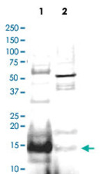
- Experimental details
- Western blot using UBE2N polyclonal antibody (Cat # PAB11286) shows detection of UBE2N protein inhuman small intestine lysate (Lane 1), but not in mouse thymus lysate (Lane 2).The heavily stained band in lane 1 (arrowhead) indicates this particular gel was overloaded with protein.The identity of minor reactive bands is unknown, but could represent E2 complexes.Each lane contains approximately 20 ug of lysate.Primary antibody was used at a 1 : 500 dilution.The membrane was washed and reacted with a 1 : 10,000 dilution of Alexa Fluor™ 680 conjugated Rb-a-Goat IgG.
