Explore
 Validate
 Learn
Learn Western blot
 Immunocytochemistry
Antibody data
- Antibody Data
- Antigen structure
- References [1]
- Comments [0]
- Validations
- Immunocytochemistry [1]
- Immunohistochemistry [1]
- Other assay [1]
Submit
Validation data
Reference
Comment
Report error
- Product number
- PA5-55172 - Provider product page

- Provider
- Invitrogen Antibodies
- Product name
- HOOK3 Polyclonal Antibody
- Antibody type
- Polyclonal
- Antigen
- Recombinant protein fragment
- Description
- Immunogen sequence: KEEIAQRCHE LDMQVAALQE EKSSLLAENQ VLMERLNQSD SIEDPNSPAG RRHLQLQTQL EQLQEETFRL EA Highest antigen sequence identity to the following orthologs: Mouse - 99%, Rat - 58%.
- Reactivity
- Human
- Host
- Rabbit
- Isotype
- IgG
- Vial size
- 100 μL
- Concentration
- 0.1 mg/mL
- Storage
- Store at 4°C short term. For long term storage, store at -20°C, avoiding freeze/thaw cycles.
Submitted references Hook3 is a scaffold for the opposite-polarity microtubule-based motors cytoplasmic dynein-1 and KIF1C.
Kendrick AA, Dickey AM, Redwine WB, Tran PT, Vaites LP, Dzieciatkowska M, Harper JW, Reck-Peterson SL
The Journal of cell biology 2019 Sep 2;218(9):2982-3001
The Journal of cell biology 2019 Sep 2;218(9):2982-3001
No comments: Submit comment
Supportive validation
- Submitted by
- Invitrogen Antibodies (provider)
- Main image
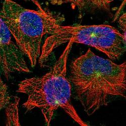
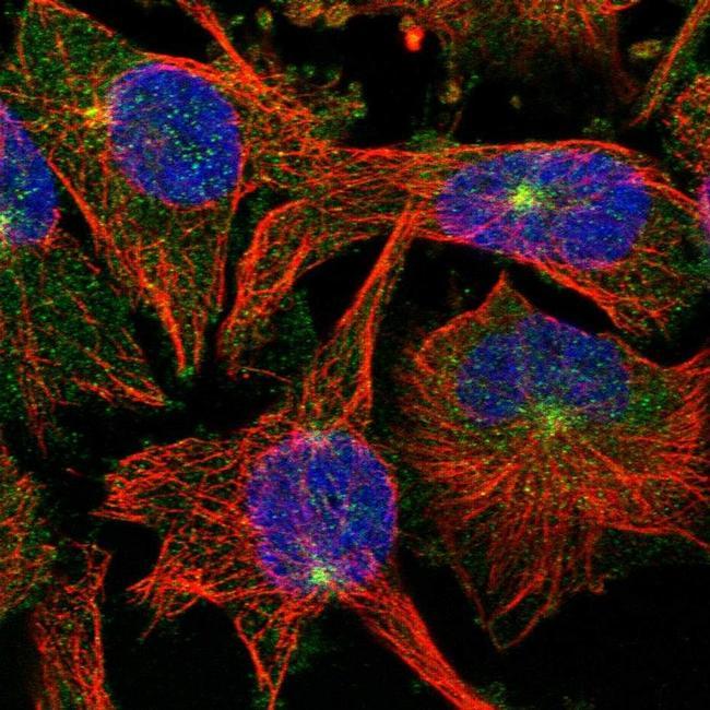
- Experimental details
- Immunofluorescent staining of HOOK3 in human cell line U-251 MG shows positivity in cytoplasm & microtubule organizing center. Samples were probed using a HOOK3 Polyclonal Antibody (Product # PA5-55172).
Supportive validation
- Submitted by
- Invitrogen Antibodies (provider)
- Main image
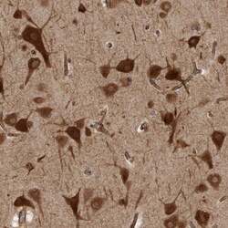
- Experimental details
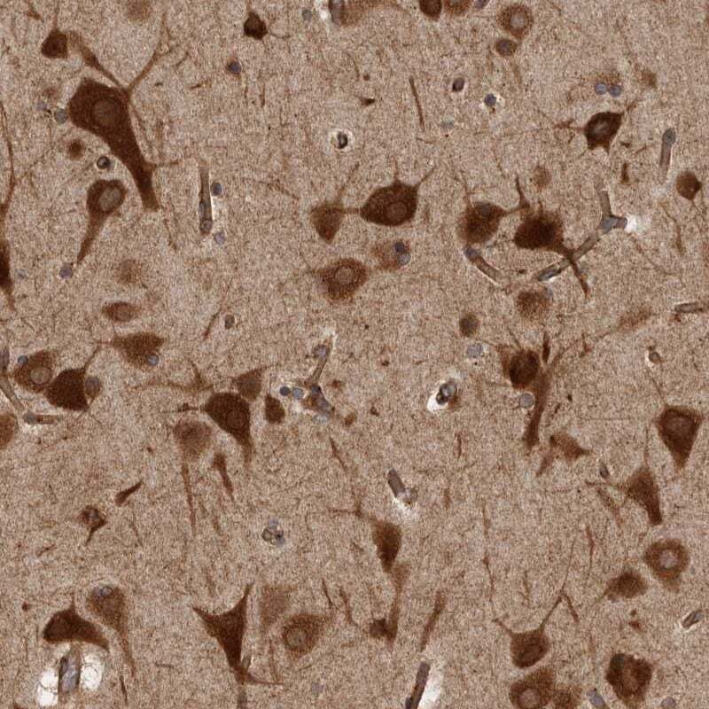
- Immunohistochemical analysis of HOOK3 in human cerebral cortex using HOOK3 Polyclonal Antibody (Product # PA5-55172) shows strong cytoplasmic positivity in neuronal cells.
Supportive validation
- Submitted by
- Invitrogen Antibodies (provider)
- Main image
- Experimental details
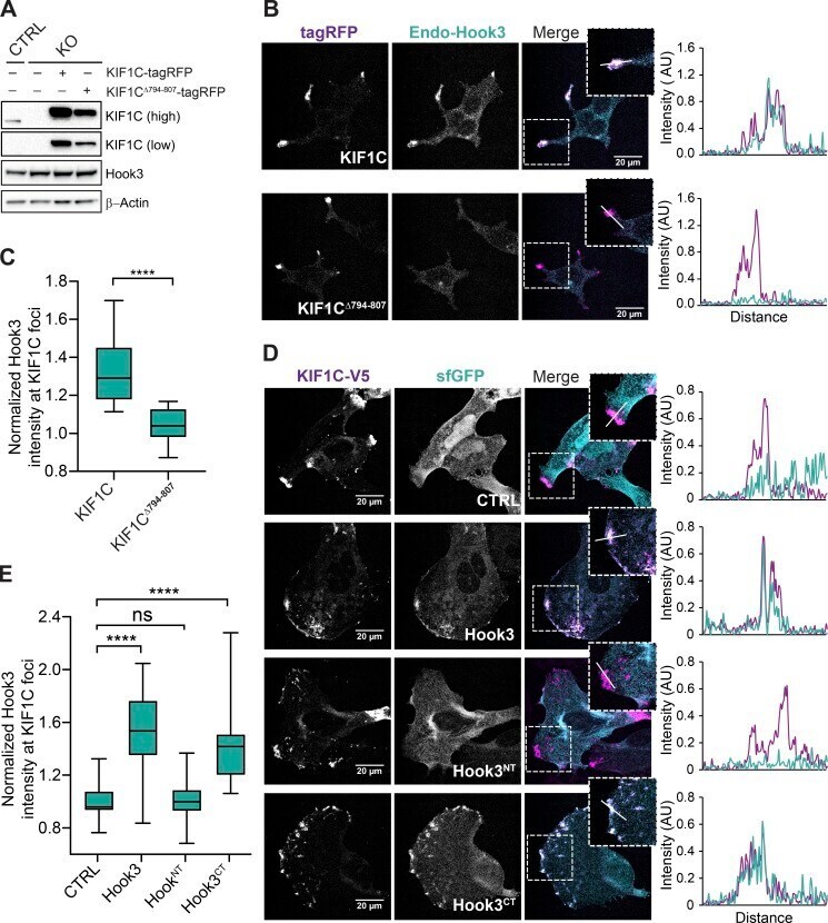
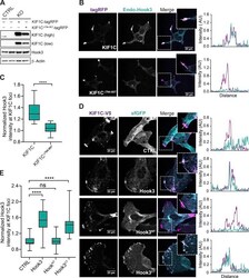
- Figure 6. KIF1C recruits Hook3 to the cell periphery. (A) 293T KIF1C KO cells (KO) were infected with viral particles encoding MSCV-driven KIF1C-tagRFP-3xFLAG or KIF1C Delta794-807 -tagRFP-3xFLAG plasmids. Immunoblots were performed with the indicated antibodies. Low and high exposures with the KIF1C antibody are shown. beta-Actin provided a loading control. 293T cells transfected with CRISPR-Cas9 (CTRL) were used as control cells. (B) Confocal microscopy of KIF1C and Hook3 localization in stable 293T cell lines expressing KIF1C-tagRFP-3xFLAG or KIF1C Delta794-807 -tagRFP-3xFLAG. Cells were grown on glass coverslips, fixed, and stained for endogenous Hook3 (Endo-Hook3). The tagRFP and Hook3 signals are shown in representative maximum intensity projections. The overlap of intensity profiles (AU) generated from drawing a 15-um line segment across individual z-sections is shown to the right of the images. (C) The mean normalized Hook3 intensity within KIF1C foci for KIF1C-tagRFP-3xFLAG ( n = 25) or KIF1C Delta794-807 -tagRFP-3xFLAG ( n = 24). Foci were determined by thresholding the KIF1C image, and masks of these foci were used to measure the Hook3 intensity in the corresponding regions in maximum projection images. Box plots represent the maximum and minimum values. Statistical significance was calculated with an unpaired t test. ****, P < 0.0001. Representative data from three independent experiments is shown. (D) Confocal microscopy of KIF1C and Hook3 in U2OS cel