Explore
 Validate
 Learn
Learn Western blot
 ELISA
ELISAAntibody data
- Antibody Data
- Antigen structure
- References [0]
- Comments [0]
- Validations
- Western blot [3]
- Immunocytochemistry [1]
Submit
Validation data
Reference
Comment
Report error
- Product number
- GTX48683 - Provider product page

- Provider
- GeneTex
- Proper citation
- GeneTex Cat#GTX48683, RRID:AB_11164441
- Product name
- RSL1D1 antibody
- Antibody type
- Polyclonal
- Reactivity
- Human
- Host
- Rabbit
No comments: Submit comment
Supportive validation
- Submitted by
- GeneTex (provider)
- Main image
- Experimental details
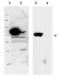
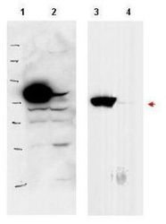
- Western blot using GeneTex's affinity purified anti-PBK1 antibody shows detection of over-expressed PBK1 in lysates from HeLa cells transfected with Flag-PBK1. Lanes 1 and 3 contain lysate from Flag-PBK1 transfected HeLa cells. Lanes 2 and 4 contain lysate from cells transfected with vector. Lanes 1 and 2 were blotted with anti-Flag antibody. Lanes 3 and 4 were probed with a 1:500 dilution of anti-PBK1. The band at 75 kDa, indicated by the arrowhead, corresponds to PBK1.
- Validation comment
- WB
- Submitted by
- GeneTex (provider)
- Main image
- Experimental details
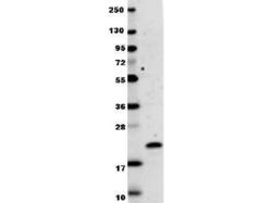
- Anti-human IL-29 antibody (GTX48683) in western blot shows detection of recombinant human IL-29 raised in E.coli. Recombinant protein (0.1 µg, 19.9 kDa) was loaded onto and resolved by SDS-PAGE, then transferred to nitrocellulose. The membrane was blocked with 1% BSA in TBST for 30 min at RT, followed by incubation with anti-Human IL-29. After washing, membrane was probed with secondary antibody Dylight 649 Conjugated Anti-Rabbit IgG (H&L) (Goat) Antibody diluted 1:20,000 in blocking buffer for 30 min. at RT.
- Validation comment
- WB

- Submitted by
- GeneTex (provider)
- Main image
- Experimental details
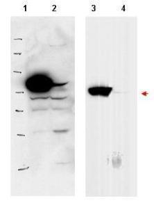
- Western blot using GeneTex's affinity purified anti-PBK1 antibody shows detection of over-expressed PBK1 in lysates from HeLa cells transfected with Flag-PBK1. Lanes 1 and 3 contain lysate from Flag-PBK1 transfected HeLa cells. Lanes 2 and 4 contain lysate from cells transfected with vector. Lanes 1 and 2 were blotted with anti-Flag antibody. Lanes 3 and 4 were probed with a 1:500 dilution of anti-PBK1. The band at 75 kDa, indicated by the arrowhead, corresponds to PBK1.

Supportive validation
- Submitted by
- GeneTex (provider)
- Main image

- Experimental details
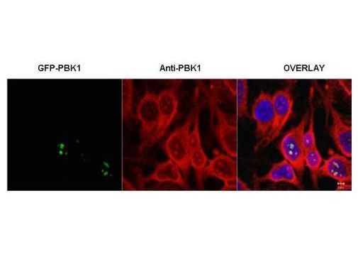
- Immunofluorescence microscopy of HeLa cells transfected with GFP-PBK1. In the overlay, specific antibody staining is shown to co-localize with recombinant protein. Cells were fixed with methanol prior to staining.