 Explore
 Validate
 Learn
Learn Western blot
Antibody data
- Antibody Data
- Antigen structure
- References [1]
- Comments [0]
- Validations
- Western blot [2]
Submit
Validation data
Reference
Comment
Report error
- Product number
- AF5359 - Provider product page

- Provider
- Novus Biologicals
- Product name
- Goat Polyclonal HABP1/C1QBP/GC1q R Antibody
- Antibody type
- Polyclonal
- Description
- Immunogen affinity purified. Detects human, mouse, and rat HABP1/C1QBP in direct ELISAs and Western blots. In direct ELISAs, less than 1% cross-reactivity with recombinant human C1QR is observed.
- Reactivity
- Human, Mouse, Rat
- Host
- Goat
- Conjugate
- Unconjugated
- Isotype
- IgG
- Vial size
- 100 ug
- Concentration
- LYOPH
- Storage
- Use a manual defrost freezer and avoid repeated freeze-thaw cycles. 12 months from date of receipt, -20 to -70 degreesC as supplied. 1 month, 2 to 8 degreesC under sterile conditions after reconstitution. 6 months, -20 to -70 degreesC under sterile conditions after reconstitution.
Submitted references Proapoptotic peptide-mediated cancer therapy targeted to cell surface p32.
Agemy L, Kotamraju VR, Friedmann-Morvinski D, Sharma S, Sugahara KN, Ruoslahti E
Molecular therapy : the journal of the American Society of Gene Therapy 2013 Dec;21(12):2195-204
Molecular therapy : the journal of the American Society of Gene Therapy 2013 Dec;21(12):2195-204
No comments: Submit comment
Supportive validation
- Submitted by
- Novus Biologicals (provider)
- Main image

- Experimental details
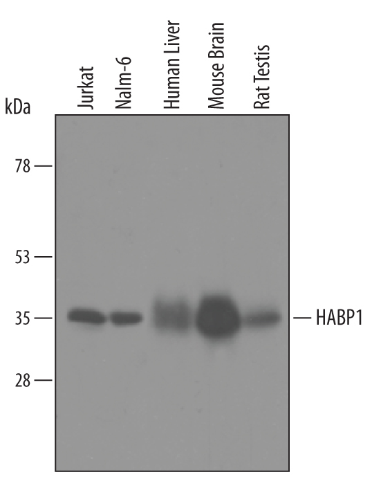
- Detection of Human/Mouse/Rat HABP1/C1QBP by Western Blot. Western blot shows lysates of Jurkat human acute T cell leukemia cell line, Nalm-6 human Pre-B acute lymphocytic leukemia cell line, human liver tissue, mouse brain tissue and rat testis tissue. PVDF membrane was probed with 1 µg/mL of Goat Anti-Human/Mouse/Rat HABP1/C1QBP Antigen Affinity-purified Polyclonal Antibody (Catalog # AF5359) followed by HRP-conjugated Anti-Goat IgG Secondary Antibody (Catalog # HAF019). A specific band was detected for HABP1/C1QBP at approximately 35 kDa (as indicated). This experiment was conducted under reducing conditions and using Immunoblot Buffer Group 8.
- Submitted by
- Novus Biologicals (provider)
- Main image

- Experimental details
- Detection of Human and Mouse HABP1/C1QBP by Simple WesternTM. Simple Western lane view shows lysates of Jurkat human acute T cell leukemia cell line and mouse brain (cortex) tissue, loaded at 0.2 mg/mL. A specific band was detected for HABP1/C1QBP at approximately 34 kDa (as indicated) using 10 µg/mL of Goat Anti-Human/Mouse/Rat HABP1/C1QBP Antigen Affinity-purified Polyclonal Antibody (Catalog # AF5359) followed by 1:50 dilution of HRP-conjugated Anti-Goat IgG Secondary Antibody (Catalog # HAF109). This experiment was conducted under reducing conditions and using the 12-230 kDa separation system.
