Explore
 Validate
 Learn
Learn Western blot
 Immunohistochemistry
Antibody data
- Antibody Data
- Antigen structure
- References [3]
- Comments [0]
- Validations
- Western blot [6]
- Immunocytochemistry [4]
- Other assay [1]
Submit
Validation data
Reference
Comment
Report error
- Product number
- PA5-35076 - Provider product page

- Provider
- Invitrogen Antibodies
- Product name
- TGFBR2 Polyclonal Antibody
- Antibody type
- Polyclonal
- Antigen
- Synthetic peptide
- Reactivity
- Human, Mouse
- Host
- Rabbit
- Isotype
- IgG
- Vial size
- 400 μL
- Concentration
- 0.5 mg/mL
- Storage
- Store at 4°C short term. For long term storage, store at -20°C, avoiding freeze/thaw cycles.
Submitted references Human Papillomavirus 16 E5 Inhibits Interferon Signaling and Supports Episomal Viral Maintenance.
POH1 contributes to hyperactivation of TGF-β signaling and facilitates hepatocellular carcinoma metastasis through deubiquitinating TGF-β receptors and caveolin-1.
Role of Dach1 revealed using a novel inner ear-specific Dach1-knockdown mouse model.
Scott ML, Woodby BL, Ulicny J, Raikhy G, Orr AW, Songock WK, Bodily JM
Journal of virology 2020 Jan 6;94(2)
Journal of virology 2020 Jan 6;94(2)
POH1 contributes to hyperactivation of TGF-β signaling and facilitates hepatocellular carcinoma metastasis through deubiquitinating TGF-β receptors and caveolin-1.
Wang B, Xu X, Yang Z, Zhang L, Liu Y, Ma A, Xu G, Tang M, Jing T, Wu L, Liu Y
EBioMedicine 2019 Mar;41:320-332
EBioMedicine 2019 Mar;41:320-332
Role of Dach1 revealed using a novel inner ear-specific Dach1-knockdown mouse model.
Miwa T, Minoda R, Ishikawa Y, Kajii T, Orita Y, Ohyama T
Biology open 2019 Aug 20;8(8)
Biology open 2019 Aug 20;8(8)
No comments: Submit comment
Supportive validation
- Submitted by
- Invitrogen Antibodies (provider)
- Main image
- Experimental details
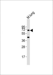
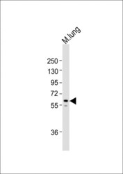
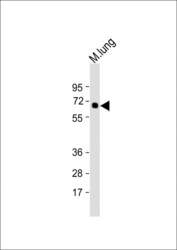
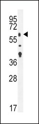
- Western blot analysis of TGFBR2 in Mouse lung lysate. Samples were incubated with TGFBR2 polyclonal antibody (Product # PA5-35076) using a dilution of 1:2,000 followed by Goat Anti-Rabbit IgG, (H+L), Peroxidase conjugated at a dilution of 1:10,000. Lysates/proteins: 20 µg per lane. Predicted band size: 65 kDa. Blocking/Dilution buffer: 5% NFDM/TBST.
- Submitted by
- Invitrogen Antibodies (provider)
- Main image
- Experimental details
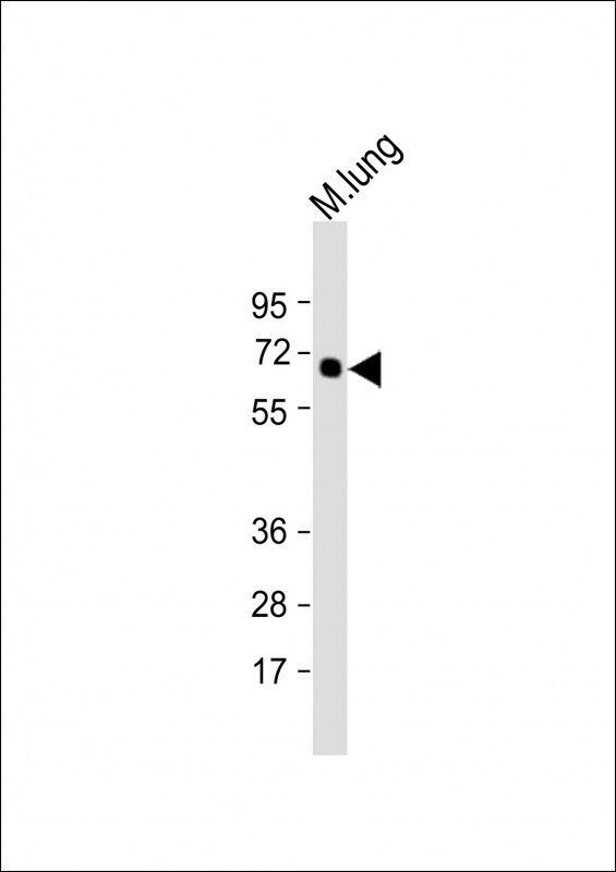
- Western blot analysis of TGFBR2 in Mouse lung lysate. Samples were incubated with TGFBR2 polyclonal antibody (Product # PA5-35076) using a dilution of 1:2,000 followed by Goat Anti-Rabbit IgG, (H+L), Peroxidase conjugated at a dilution of 1:10,000. Lysates/proteins: 20 µg per lane. Predicted band size: 67 kDa. Blocking/Dilution buffer: 5% NFDM/TBST.
- Submitted by
- Invitrogen Antibodies (provider)
- Main image
- Experimental details
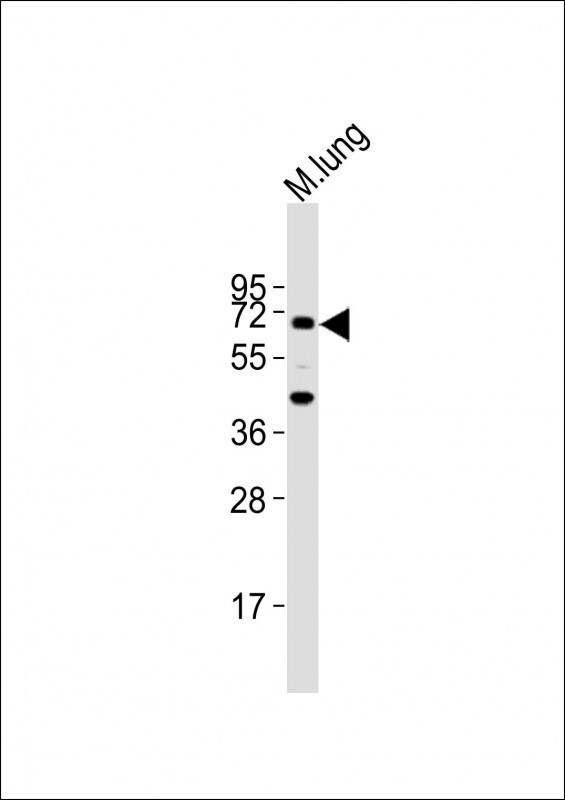
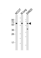
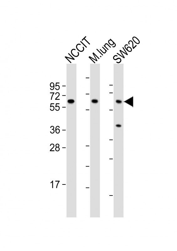
- Western blot analysis of TGFBR2 in various lysates. Samples were incubated with TGFBR2 polyclonal antibody (Product # PA5-35076) using a dilution of 1:2,000 followed by Goat Anti-Rabbit IgG, (H+L), Peroxidase conjugated at a dilution of 1:10,000. Lysates/proteins: 20 µg per lane. Lane 1: NCCIT whole cell lysates; Lane 2: mouse lung lysates; Lane 3: SW620 whole cell lysates. Predicted band size: 67 kDa. Blocking/Dilution buffer: 5% NFDM/TBST.
- Submitted by
- Invitrogen Antibodies (provider)
- Main image

- Experimental details
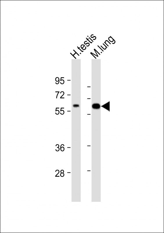
- Western blot analysis of TGFBR2 in various lysates. Samples were incubated with TGFBR2 polyclonal antibody (Product # PA5-35076) using a dilution of 1:2,000 followed by Goat Anti-Rabbit IgG, (H+L), Peroxidase conjugated at a dilution of 1:10,000. Lysates/proteins: 20 µg per lane. Lane 1: human testis lysate; Lane 2: mouse lung lysate. Predicted band size: 67 kDa. Blocking/Dilution buffer: 5% NFDM/TBST.
- Submitted by
- Invitrogen Antibodies (provider)
- Main image
- Experimental details
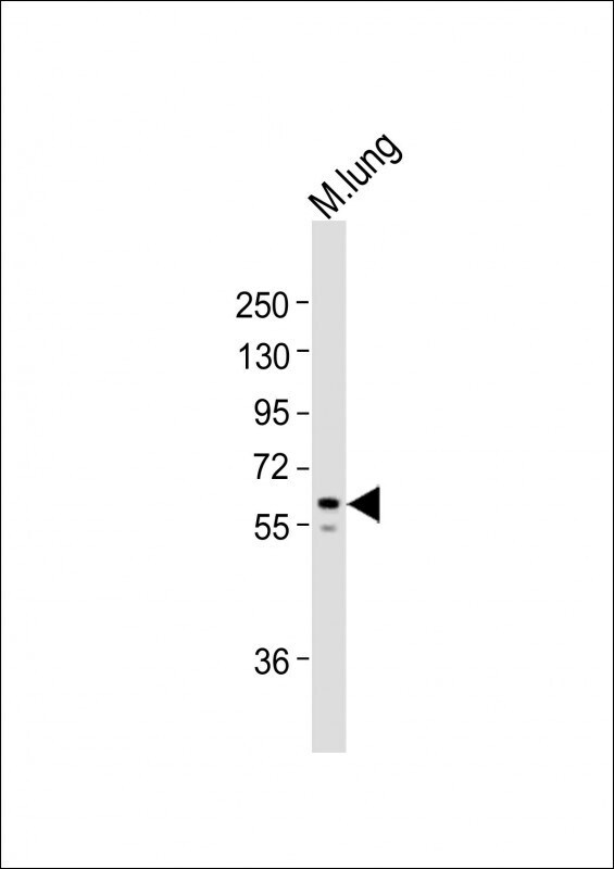
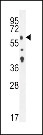
- Western blot analysis of TGFBR2 in mouse lung tissue lysates. Samples were incubated with TGFBR2 polyclonal antibody (Product # PA5-35076). Lysates: 35 µg/lane. TGFBR2 protein (arrow).
Supportive validation
- Submitted by
- Invitrogen Antibodies (provider)
- Main image

- Experimental details
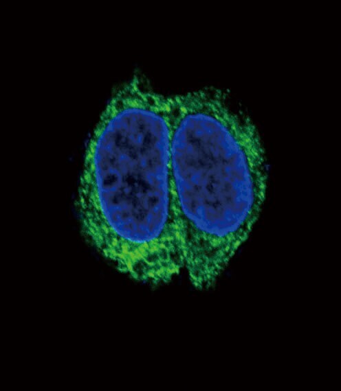
- Immunofluorescent analysis of TGFBR2 in HepG2 cells using a TGFBR2 polyclonal antibody (Product # PA5-35076) followed by detection using a fluorescent conjugated secondary antibody (green). Nuclei were stained with Dapi (blue).

- Submitted by
- Invitrogen Antibodies (provider)
- Main image

- Experimental details
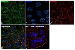
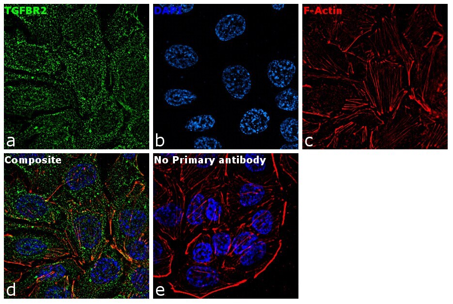
- Immunofluorescence analysis of TGFBR2 was performed using 70% confluent log phase HT-29 cells. The cells were fixed with 4% paraformaldehyde for 10 minutes, permeabilized with 0.1% Triton™ X-100 for 15 minutes, and blocked with 1% BSA for 1 hour at room temperature. The cells were labeled with TGFBR2 Rabbit Polyclonal Antibody(Product # PA5-35076) at 1:100 dilution in 0.1% BSA, incubated at 4 degree Celsius overnight and then labeled with Goat anti-Rabbit IgG (H+L) Superclonal™ Secondary Antibody, Alexa Fluor® 488 conjugate (Product # A27034) at a dilution of 1:2000 for 45 minutes at room temperature (Panel a: green). Nuclei (Panel b: blue) were stained with ProLong™ Diamond Antifade Mountant with DAPI (Product # P36962). F-actin (Panel c: red) was stained with Rhodamine Phalloidin (Product # R415, 1:300). Panel d represents the merged image showing membrane and cytoplasmic localization. Panel e represents control cells with no primary antibody to assess background. The images were captured at 60X magnification.

- Submitted by
- Invitrogen Antibodies (provider)
- Main image
- Experimental details
- Immunofluorescence analysis of TGFBR2 was performed using 70% confluent log phase HT-29 cells. The cells were fixed with 4% paraformaldehyde for 10 minutes, permeabilized with 0.1% Triton™ X-100 for 15 minutes, and blocked with 1% BSA for 1 hour at room temperature. The cells were labeled with TGFBR2 Rabbit Polyclonal Antibody(Product # PA5-35076) at 1:100 dilution in 0.1% BSA, incubated at 4 degree Celsius overnight and then labeled with Goat anti-Rabbit IgG (Heavy Chain) Superclonal™ Secondary Antibody, Alexa Fluor® 488 conjugate (Product # A27034) at a dilution of 1:2000 for 45 minutes at room temperature (Panel a: green). Nuclei (Panel b: blue) were stained with ProLong™ Diamond Antifade Mountant with DAPI (Product # P36962). F-actin (Panel c: red) was stained with Rhodamine Phalloidin (Product # R415, 1:300). Panel d represents the merged image showing membrane and cytoplasmic localization. Panel e represents control cells with no primary antibody to assess background. The images were captured at 60X magnification.
- Submitted by
- Invitrogen Antibodies (provider)
- Main image

- Experimental details
- Immunocytochemistry analysis of TGFBR2 in HepG2 cells. Samples were incubated in TGFBR2 polyclonal antibody (Product # PA5-35076) followed by Alexa Fluor 488-conjugated goat anti-rabbit lgG (green). DAPI was used to stain the cell nuclear (blue).
Supportive validation
- Submitted by
- Invitrogen Antibodies (provider)
- Main image

- Experimental details
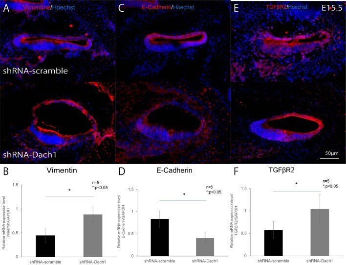
- Fig. 5. The EMT in the lateral wall of the treated developing cochlear duct. (A-F) Protein and mRNA expression in the lateral wall of the cochlear epithelium, utilising immunohistochemistry and qRT-PCR analyses in inner ear-specific Dach1-KD mouse embryos and control embryos at E15.5. (A,B) Vimentin (* P =0.03) (C,D) E-cadherin (* P =0.04). (E,F) TGFbetaR2 (* P =0.04).