Explore
 Validate
 Learn
LearnHPA018904
antibody from Atlas Antibodies
Targeting: SPEG
APEG1, BPEG, KIAA1297, MGC12676, MYLK6, SPEGalpha, SPEGbeta
 Immunohistochemistry
Antibody data
- Antibody Data
- Antigen structure
- References [1]
- Comments [0]
- Validations
- Immunohistochemistry [1]
Submit
Validation data
Reference
Comment
Report error
- Product number
- HPA018904 - Provider product page

- Provider
- Atlas Antibodies
- Proper citation
- Atlas Antibodies Cat#HPA018904, RRID:AB_1844924
- Product name
- Anti-SPEG
- Antibody type
- Polyclonal
- Description
- Polyclonal Antibody against Human SPEG, Gene description: SPEG complex locus, Alternative Gene Names: APEG1, BPEG, KIAA1297, MGC12676, SPEGalpha, SPEGbeta, Validated applications: IHC, Uniprot ID: Q15772, Storage: Store at +4°C for short term storage. Long time storage is recommended at -20°C.
- Reactivity
- Human
- Host
- Rabbit
- Conjugate
- Unconjugated
- Isotype
- IgG
- Vial size
- 100 µl
- Concentration
- 0.2 mg/ml
- Storage
- Store at +4°C for short term storage. Long time storage is recommended at -20°C.
- Handling
- The antibody solution should be gently mixed before use.
Submitted references Striated muscle-specific serine/threonine-protein kinase beta segregates with high versus low responsiveness to endurance exercise training
Kusić D, Connolly J, Kainulainen H, Semenova E, Borisov O, Larin A, Popov D, Generozov E, Ahmetov I, Britton S, Koch L, Burniston J
Physiological Genomics 2020;52(1):35-46
Physiological Genomics 2020;52(1):35-46
No comments: Submit comment
Supportive validation
- Submitted by
- Atlas Antibodies (provider)
- Enhanced method
- Orthogonal validation
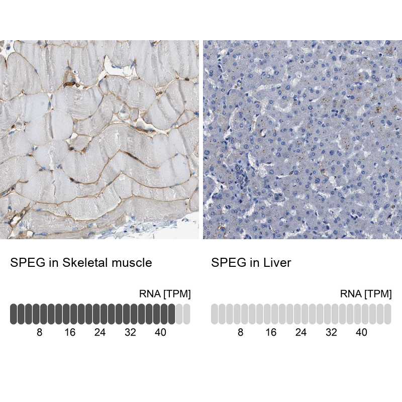
- Main image
- Experimental details
- Immunohistochemistry analysis in human skeletal muscle and liver tissues using HPA018904 antibody. Corresponding SPEG RNA-seq data are presented for the same tissues.
- Sample type
- Human
- Protocol
- Protocol