 Explore
 Validate
 Learn
Learn Western blot
 Immunoprecipitation
Antibody data
- Antibody Data
- Antigen structure
- References [0]
- Comments [0]
- Validations
- Western blot [3]
- Flow cytometry [6]
Submit
Validation data
Reference
Comment
Report error
- Product number
- MA1-19285 - Provider product page

- Provider
- Invitrogen Antibodies
- Product name
- SIT Monoclonal Antibody (SIT-01)
- Antibody type
- Monoclonal
- Antigen
- Recombinant protein fragment
- Description
- This antibody reacts with murine SIT. Western Blot: Reducing conditions.
- Reactivity
- Human
- Host
- Mouse
- Isotype
- IgG
- Antibody clone number
- SIT-01
- Vial size
- 100 μg
- Concentration
- 1 mg/mL
- Storage
- 4°C, do not freeze
No comments: Submit comment
Supportive validation
- Submitted by
- Invitrogen Antibodies (provider)
- Main image
- Experimental details
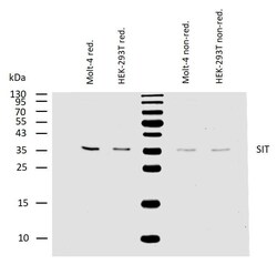
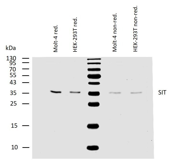
- Western blotting analysis of human SIT using mouse monoclonal antibody SIT-01 on lysates of Molt-4 and HEK-293T cells under reducing and non-reducing conditions. Nitrocellulose membrane was probed with 2µg/mL of mouse anti-SIT Monoclonal antibody (Product # MA1-192850) followed by IRDye800-conjugated anti-mouse secondary antibody. SIT was detected around 36kDa.
- Submitted by
- Invitrogen Antibodies (provider)
- Main image
- Experimental details
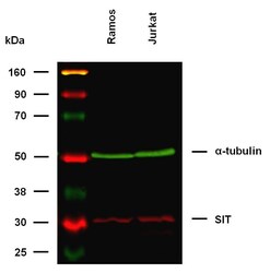
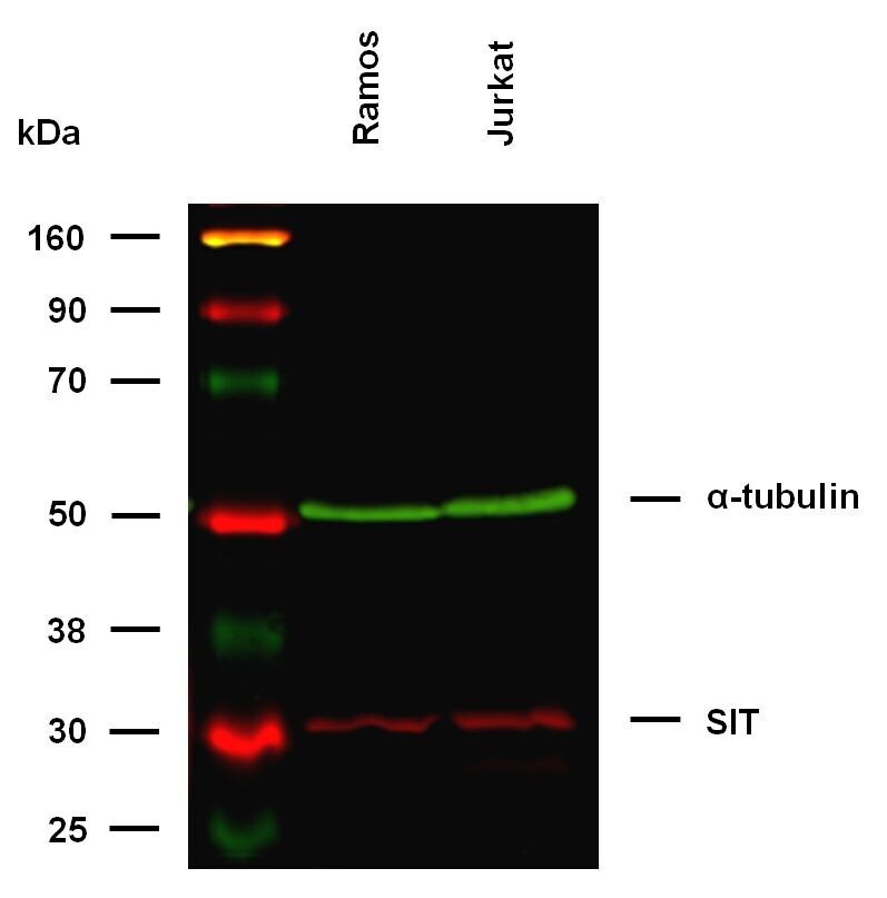
- Western Blot analysis of SIT using SIT Monoclonal Antibody (SIT-01) (Product # MA1-19285). Analysis was performed on whole cell extracts (RIPA lysis buffer) of Ramos and Jurkat cell lines, mixed and heated (100°C, 5 min) with reducing (2-mercaptoethanol) SDS-loading buffer. Samples were resolved using 10% Tris-glycine SDS gel electrophoresis. Nitrocellulose membrane blot was probed simultaneously with mouse IgG1 monoclonal antibody SIT-01 (2 µg/mL), and rat IgG2a anti-tubulin monoclonal antibody (1 µg/mL) used as the loading control. Subclass-specific secondary antibodies Goat-anti-Rat IgG (green) and Goat-anti-Mouse IgG (red) were used for multiplex fluorescent Western blot detection. SIT was detected at ~32 kDa in tested cell lines.
- Submitted by
- Invitrogen Antibodies (provider)
- Main image
- Experimental details
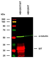
- Western Blot analysis of SIT using SIT Monoclonal Antibody (SIT-01) (Product # MA1-19285). The specificity of SIT-01 antibody was assessed by comparing binding signals in HEK293T cells overexpressing the target SIT protein to wild type cells (control) with low level of endogenous protein expression. Western blotting analysis was performed on whole cell extracts (urea lysis buffer) of transfected and control cells, mixed and heated (100°C, 5 min) with reducing (2-mercaptoethanol) SDS-loading buffer. Samples were resolved using 10% Tris-glycine SDS gel electrophoresis. Nitrocellulose membrane blot was probed simultaneously with mouse IgG1 monoclonal antibody (2 µg/mL), and rat IgG2a anti-tubulin monoclonal antibody (1 µg/mL) used as the loading control. Subclass-specific secondary antibodies Goat-anti-Rat IgG (green) and Goat-anti-Mouse IgG (red) were used for multiplex fluorescent Western blot detection.

Supportive validation
- Submitted by
- Invitrogen Antibodies (provider)
- Main image
- Experimental details
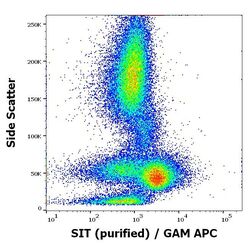
- Flow cytometry intracellular staining pattern of human peripheral whole blood using anti-SIT (SIT-01) purified Monoclonal antibody (Product # MA1-19285) (concentration in sample 9 µg/mL, GAM APC).
- Submitted by
- Invitrogen Antibodies (provider)
- Main image

- Experimental details
- Separation of human CD3 negative SIT positive lymphocytes (red-filled) from CD3 negative SIT negative lymphocytes (black-dashed) in flow cytometry analysis (intracellular staining) of peripheral human whole blood stained using anti-SIT (SIT-01) purified Monoclonal antibody (Product # MA1-19285) (concentration in sample 9 µg/mL, GAM APC).

- Submitted by
- Invitrogen Antibodies (provider)
- Main image

- Experimental details
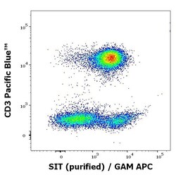
- Flow cytometry multicolor intracellular staining of human peripheral whole blood stained using anti-SIT (SIT-01) purified Monoclonal antibody (Product # MA1-19285) (concentration in sample 9 µg/mL, GAM APC) and anti-human CD3 (UCHT1) Pacific Blue™ using a dilution of 20 µL reagent/100 µL of peripheral whole blood.

- Submitted by
- Invitrogen Antibodies (provider)
- Main image

- Experimental details
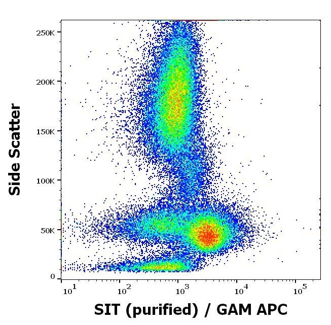
- Flow cytometry intracellular staining pattern of human peripheral whole blood using anti-SIT (SIT-01) purified Monoclonal antibody (Product # MA1-19285) (concentration in sample 9 µg/mL, GAM APC).

- Submitted by
- Invitrogen Antibodies (provider)
- Main image
- Experimental details
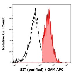
- Separation of human CD3 negative SIT positive lymphocytes (red-filled) from CD3 negative SIT negative lymphocytes (black-dashed) in flow cytometry analysis (intracellular staining) of peripheral human whole blood stained using anti-SIT (SIT-01) purified Monoclonal antibody (Product # MA1-19285) (concentration in sample 9 µg/mL, GAM APC).

- Submitted by
- Invitrogen Antibodies (provider)
- Main image
- Experimental details
- Flow cytometry multicolor intracellular staining of human peripheral whole blood stained using anti-SIT (SIT-01) purified Monoclonal antibody (Product # MA1-19285) (concentration in sample 9 µg/mL, GAM APC) and anti-human CD3 (UCHT1) Pacific Blue™ using a dilution of 20 µL reagent/100 µL of peripheral whole blood.
