Explore
 Validate
 Learn
Learn Western blot
Antibody data
- Antibody Data
- Antigen structure
- References [0]
- Comments [0]
- Validations
- Western blot [4]
- Immunocytochemistry [1]
Submit
Validation data
Reference
Comment
Report error
- Product number
- 711811 - Provider product page

- Provider
- Invitrogen Antibodies
- Product name
- SPG11 Recombinant Superclonal™ Antibody (2HCLC)
- Antibody type
- Other
- Antigen
- Synthetic peptide
- Description
- This antibody is predicted to react with Monkey, Horse, Bovine, Sheep Recombinant rabbit Superclonal™ antibodies are unique offerings from Thermo Fisher Scientific. They are comprised of a selection of multiple different recombinant monoclonal antibodies, providing the best of both worlds - the sensitivity of polyclonal antibodies with the specificity of monoclonal antibodies - all delivered with the consistency only found in a recombinant antibody. While functionally the same as a polyclonal antibody - recognizing multiple epitope sites on the target and producing higher detection sensitivity for low abundance targets - a recombinant rabbit Superclonal™ antibody has a known mixture of light and heavy chains. The exact population can be produced in every lot, circumventing the biological variability typically associated with polyclonal antibody production. Note: Formerly called “Recombinant polyclonal antibody”, this product is now rebranded as “Recombinant Superclonal™ antibody”. The physical product and the performance remain unchanged.
- Reactivity
- Human
- Host
- Rabbit
- Isotype
- IgG
- Antibody clone number
- 2HCLC
- Vial size
- 100 μg
- Concentration
- 0.5 mg/mL
- Storage
- Store at 4°C short term. For long term storage, store at -20°C, avoiding freeze/thaw cycles.
No comments: Submit comment
Supportive validation
- Submitted by
- Invitrogen Antibodies (provider)
- Main image

- Experimental details
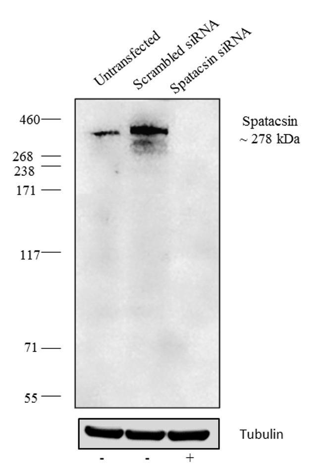
- Knockdown of Spatacsin was achieved by transfecting HeLa cells with Spatacsin specific validated siRNA (Silencer® select Product # s37058 and s37059). Western blot analysis was performed using membrane extracts from the Spatacsin knock down cells (lane 3), non-specific scrambled siRNA transfected cells (lane 2) and untransfected cells (lane 1). The blots were probed with Anti-Spatacsin Recombinant Rabbit Superclonal™ Antibody (Product # 711811, 1-3 µg/mL) and Goat anti-Rabbit IgG (Heavy Chain) Superclonal™ Secondary Antibody, HRP conjugate (Product # A27036, 0.4 µg/mL, 1:2500 dilution). Loss of signal upon siRNA mediated knock down confirms that antibody is specific to Spatacsin.
- Submitted by
- Invitrogen Antibodies (provider)
- Main image

- Experimental details
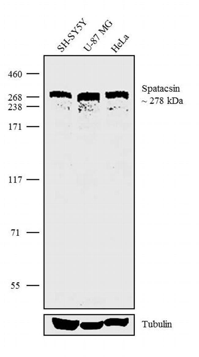
- Western blot analysis was performed on Whole cell extracts (30 µg lysate) of SH-SY5Y (Lane 1), U-87 MG (Lane 2) and HeLa (Lane 3). The blots were probed with Anti-Spatacsin Recombinant Rabbit Superclonal™ Antibody (Product # 711811, 2.5 µg/mL) and detected by chemiluminescence using Goat anti-Rabbit IgG (Heavy Chain) Superclonal™ Secondary Antibody, HRP conjugate (Product # A27036, 0.4 µg/mL, 1:4000 dilution). A 278 kDa band corresponding to Spatacsin was observed across the cell lines tested. Known quantity of protein samples were electrophoresed using Novex®NuPAGE®4-12% Bis-Tris gel (Product # NP0322BOX), XCell SureLock™ Electrophoresis System (Product # EI0002) and HiMark™ Pre-Stained Protein Standard (Product # LC5699). Resolved proteins were then transferred onto a nitrocellulose membrane by wet transfer method. The membrane was probed with the relevant primary and secondary Antibody following blocking with 5% skimmed milk. Chemiluminescent detection was performed using Pierce™ ECL Western blotting Substrate (Product # 32106).
- Submitted by
- Invitrogen Antibodies (provider)
- Main image
- Experimental details
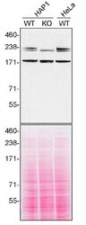
- Western blot of SPG11 was performed by loading 30 µg of HAP1 WT (lane 1) and SPG11 KO (lane 2) cell lysates in RIPA buffer onto a NuPAGE™ 3 to 8%, Tris-Acetate, 1.0 mm, Midi Protein Gel (Product # WG1601BOX). Proteins on the blot were visualized with Ponceau staining (below immunoblot). Proteins were transferred to nitrocellulose membrane and blocked in 5% milk for 1 hr. SPG11 was detected at approximately 279 kDa using SPG11 Recombinant Rabbit Superclonal™ Antibody (2HCLC) (Product # 711811) at a dilution of 1:200 in 5% BSA in TBST overnight at 4 degree celsius. The blot was probed with Goat anti-Rabbit IgG (H+L) Secondary Antibody, HRP (Product # 65-6120) diluted to 0.2 µg/mL in TBST with 5% milk for 1 hr at room temperature. Chemiluminescent detection was performed using ECL Western Blotting Substrate. Data courtesy of YCharOS Inc., an open science company with the mission of characterizing commercially available antibodies using knockout validation.

- Submitted by
- Invitrogen Antibodies (provider)
- Main image
- Experimental details
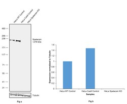
- Knockout of Spatacsin was achieved by CRISPR-Cas9 genome editing using LentiArray™ Lentiviral sgRNA (Product # A32042, Assay ID CRISPR742574_LV) and LentiArray Cas9 Lentivirus (Product # A32064). Western blot analysis of Spatacsin was performed by loading 30 µg of HeLa wild type (Lane 1), HeLa Cas9 (Lane 2) and HeLa Spatacsin KO (Lane 3) whole cell extracts. The samples were electrophoresed using NuPAGE™ 3 to 8%, Tris-Acetate, 1.0 mm, Mini Protein Gel (Product # EC6695BOX). Resolved proteins were then transferred onto a nitrocellulose membrane (Product # IB23001) by iBlot® 2 Dry Blotting System (Product # IB21001). The blot was probed with SPG11 Recombinant Rabbit Superclonal™ Antibody (2HCLC) (Product # 711811, 1:200 dilution) and detected by Goat anti-Rabbit IgG (H+L) Superclonal™ Recombinant Secondary Antibody, HRP (Product # A27036, 1:10,000 dilution) using the iBright™ FL1500 (Product # A44115). Chemiluminescent detection was performed using SuperSignal™ West Dura Extended Duration Substrate (Product # 34076). Loss of signal upon CRISPR mediated knockout (KO) using the LentiArray™ CRISPR product line confirms that antibody is specific to Spatacsin.
Supportive validation
- Submitted by
- Invitrogen Antibodies (provider)
- Main image
- Experimental details
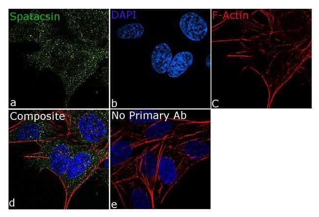
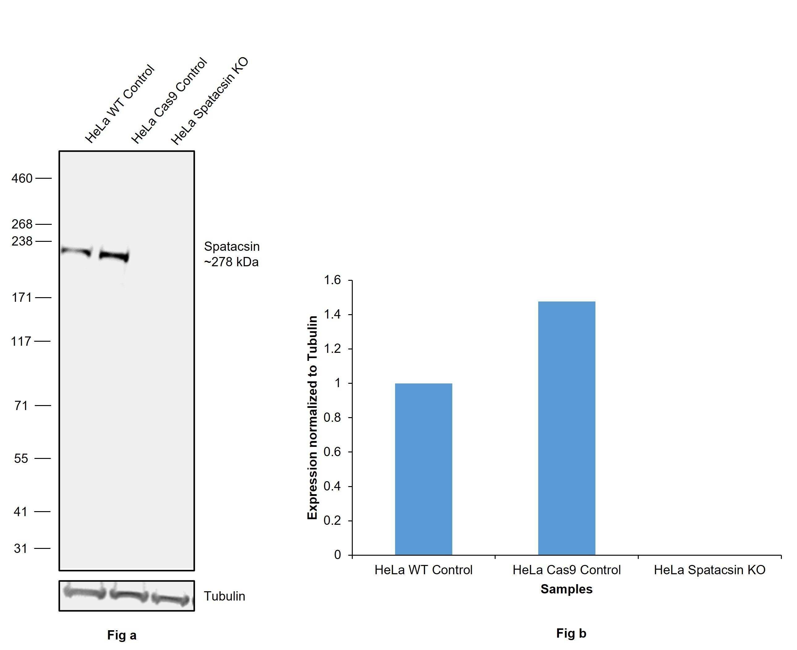
- For immunofluorescence analysis, SH-SY5Y cells were fixed and permeabilized for detection of endogenous Spatacsin using Anti- Spatacsin Recombinant Rabbit Superclonal™ Antibody (Product # 711811, 5 µg/mL) and labeled with Goat anti-Rabbit IgG (Heavy Chain) Superclonal™ Secondary Antibody, Alexa Fluor® 488 conjugate (Product # A27034, 1:2000). Panel a) shows representative cells that were stained for detection and localization of Spatacsin (green), Panel b) is stained for nuclei (blue) using SlowFade® Gold Antifade Mountant with DAPI (Product # S36938). Panel c) represents cytoskeletal F-actin staining using Rhodamine Phalloidin (Product # R415, 1:300). Panel d) is a composite image of Panels a, b and c clearly demonstrating cytoplasmic localization of Spatacsin. Panel e) represents control cells with no primary antibody to assess background. The images were captured at 60X magnification.