 Explore
 Validate
 Learn
Learn Western blot
 Immunohistochemistry
Antibody data
- Antibody Data
- Antigen structure
- References [9]
- Comments [0]
- Validations
- Western blot [2]
Submit
Validation data
Reference
Comment
Report error
- Product number
- AF2147 - Provider product page

- Provider
- R&D Systems
- Product name
- Mouse CHL-1/L1CAM-2 Antibody
- Antibody type
- Polyclonal
- Description
- Antigen Affinity-purified. Detects mouse CHL-1/L1CAM-2 in direct ELISAs and Western blots. In direct ELISAs and Western blots, approximately 40% cross-reactivity with recombinant human CHL-1 is observed.
- Reactivity
- Mouse
- Host
- Goat
- Conjugate
- Unconjugated
- Antigen sequence
BAC30699- Isotype
- IgG
- Vial size
- 100 ug
- Concentration
- LYOPH
- Storage
- Use a manual defrost freezer and avoid repeated freeze-thaw cycles. 12 months from date of receipt, -20 to -70 °C as supplied. 1 month, 2 to 8 °C under sterile conditions after reconstitution. 6 months, -20 to -70 °C under sterile conditions after reconstitution.
Submitted references BACE1 elevation engendered by GGA3 deletion increases β-amyloid pathology in association with APP elevation and decreased CHL1 processing in 5XFAD mice.
An aberrant sugar modification of BACE1 blocks its lysosomal targeting in Alzheimer's disease.
Transcriptome analysis reveals transmembrane targets on transplantable midbrain dopamine progenitors.
β-Site amyloid precursor protein (APP)-cleaving enzyme 1 (BACE1)-deficient mice exhibit a close homolog of L1 (CHL1) loss-of-function phenotype involving axon guidance defects.
EphB regulates L1 phosphorylation during retinocollicular mapping.
Secretome protein enrichment identifies physiological BACE1 protease substrates in neurons.
CHL1 negatively regulates the proliferation and neuronal differentiation of neural progenitor cells through activation of the ERK1/2 MAPK pathway.
Phosphatidylinositol 3-kinase/protein kinase Cdelta activation induces close homolog of adhesion molecule L1 (CHL1) expression in cultured astrocytes.
CHL1 promotes Sema3A-induced growth cone collapse and neurite elaboration through a motif required for recruitment of ERM proteins to the plasma membrane.
Kim W, Ma L, Lomoio S, Willen R, Lombardo S, Dong J, Haydon PG, Tesco G
Molecular neurodegeneration 2018 Feb 2;13(1):6
Molecular neurodegeneration 2018 Feb 2;13(1):6
An aberrant sugar modification of BACE1 blocks its lysosomal targeting in Alzheimer's disease.
Kizuka Y, Kitazume S, Fujinawa R, Saito T, Iwata N, Saido TC, Nakano M, Yamaguchi Y, Hashimoto Y, Staufenbiel M, Hatsuta H, Murayama S, Manya H, Endo T, Taniguchi N
EMBO molecular medicine 2015 Feb;7(2):175-89
EMBO molecular medicine 2015 Feb;7(2):175-89
Transcriptome analysis reveals transmembrane targets on transplantable midbrain dopamine progenitors.
Bye CR, Jönsson ME, Björklund A, Parish CL, Thompson LH
Proceedings of the National Academy of Sciences of the United States of America 2015 Apr 14;112(15):E1946-55
Proceedings of the National Academy of Sciences of the United States of America 2015 Apr 14;112(15):E1946-55
β-Site amyloid precursor protein (APP)-cleaving enzyme 1 (BACE1)-deficient mice exhibit a close homolog of L1 (CHL1) loss-of-function phenotype involving axon guidance defects.
Hitt B, Riordan SM, Kukreja L, Eimer WA, Rajapaksha TW, Vassar R
The Journal of biological chemistry 2012 Nov 9;287(46):38408-25
The Journal of biological chemistry 2012 Nov 9;287(46):38408-25
EphB regulates L1 phosphorylation during retinocollicular mapping.
Dai J, Dalal JS, Thakar S, Henkemeyer M, Lemmon VP, Harunaga JS, Schlatter MC, Buhusi M, Maness PF
Molecular and cellular neurosciences 2012 Jun;50(2):201-10
Molecular and cellular neurosciences 2012 Jun;50(2):201-10
Secretome protein enrichment identifies physiological BACE1 protease substrates in neurons.
Kuhn PH, Koroniak K, Hogl S, Colombo A, Zeitschel U, Willem M, Volbracht C, Schepers U, Imhof A, Hoffmeister A, Haass C, Roßner S, Bräse S, Lichtenthaler SF
The EMBO journal 2012 Jun 22;31(14):3157-68
The EMBO journal 2012 Jun 22;31(14):3157-68
CHL1 negatively regulates the proliferation and neuronal differentiation of neural progenitor cells through activation of the ERK1/2 MAPK pathway.
Huang X, Zhu LL, Zhao T, Wu LY, Wu KW, Schachner M, Xiao ZC, Fan M
Molecular and cellular neurosciences 2011 Jan;46(1):296-307
Molecular and cellular neurosciences 2011 Jan;46(1):296-307
Phosphatidylinositol 3-kinase/protein kinase Cdelta activation induces close homolog of adhesion molecule L1 (CHL1) expression in cultured astrocytes.
Wu J, Wrathall JR, Schachner M
Glia 2010 Feb;58(3):315-28
Glia 2010 Feb;58(3):315-28
CHL1 promotes Sema3A-induced growth cone collapse and neurite elaboration through a motif required for recruitment of ERM proteins to the plasma membrane.
Schlatter MC, Buhusi M, Wright AG, Maness PF
Journal of neurochemistry 2008 Feb;104(3):731-44
Journal of neurochemistry 2008 Feb;104(3):731-44
No comments: Submit comment
Supportive validation
- Submitted by
- R&D Systems (provider)
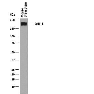
- Main image
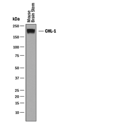
- Experimental details
- Detection of Mouse CHL-1/L1CAM-2 by Western Blot. Western blot shows lysates of mouse brain stem tissue. PVDF membrane was probed with 0.25 µg/mL of Goat Anti-Mouse CHL-1/L1CAM-2 Antigen Affinity-purified Polyclonal Antibody (Catalog # AF2147) followed by HRP-conjugated Anti-Goat IgG Secondary Antibody (Catalog # HAF019). A specific band was detected for CHL-1/L1CAM-2 at approximately 200 kDa (as indicated). This experiment was conducted under reducing conditions and using Immunoblot Buffer Group 1.
- Submitted by
- R&D Systems (provider)
- Main image
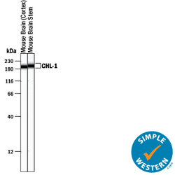
- Experimental details
- Detection of Mouse CHL-1/L1CAM-2 by Simple WesternTM. Simple Western lane view shows lysates of mouse brain (cortex) tissue and mouse brain stem tissue, loaded at 0.2 mg/mL. A specific band was detected for CHL-1/L1CAM-2 at approximately 196-201 kDa (as indicated) using 2.5 µg/mL of Goat Anti-Mouse CHL-1/L1CAM-2 Antigen Affinity-purified Polyclonal Antibody (Catalog # AF2147) followed by 1:50 dilution of HRP-conjugated Anti-Goat IgG Secondary Antibody (Catalog # HAF109) . This experiment was conducted under reducing conditions and using the 12-230 kDa separation system.
