Explore
 Validate
 Learn
Learn Western blot
 Immunoprecipitation
Antibody data
- Antibody Data
- Antigen structure
- References [5]
- Comments [0]
- Validations
- Western blot [1]
Submit
Validation data
Reference
Comment
Report error
- Product number
- MAB1934 - Provider product page

- Provider
- R&D Systems
- Product name
- Human DEP-1/CD148 Antibody
- Antibody type
- Monoclonal
- Description
- Protein A or G purified from hybridoma culture supernatant. Detects human DEP-1/CD148 in Western blots.
- Reactivity
- Human
- Host
- Mouse
- Conjugate
- Unconjugated
- Isotype
- IgG
- Antibody clone number
- 143-41
- Vial size
- 100 ug
- Concentration
- LYOPH
- Storage
- Use a manual defrost freezer and avoid repeated freeze-thaw cycles. 12 months from date of receipt, -20 to -70 °C as supplied. 1 month, 2 to 8 °C under sterile conditions after reconstitution. 6 months, -20 to -70 °C under sterile conditions after reconstitution.
Submitted references A novel splice variant of the protein tyrosine phosphatase PTPRJ that encodes for a soluble protein involved in angiogenesis.
The protein tyrosine phosphatase DEP-1/PTPRJ promotes breast cancer cell invasion and metastasis.
Phosphorylation of DEP-1/PTPRJ on threonine 1318 regulates Src activation and endothelial cell permeability induced by vascular endothelial growth factor.
Systematic validation of specific phenotypic markers for in vitro polarized human macrophages.
Analysis of HLDA9 mAbs on plasmacytoid dendritic cells.
Bilotta A, Dattilo V, D'Agostino S, Belviso S, Scalise S, Bilotta M, Gaudio E, Paduano F, Perrotti N, Florio T, Fusco A, Iuliano R, Trapasso F
Oncotarget 2017 Feb 7;8(6):10091-10102
Oncotarget 2017 Feb 7;8(6):10091-10102
The protein tyrosine phosphatase DEP-1/PTPRJ promotes breast cancer cell invasion and metastasis.
Spring K, Fournier P, Lapointe L, Chabot C, Roussy J, Pommey S, Stagg J, Royal I
Oncogene 2015 Oct 29;34(44):5536-47
Oncogene 2015 Oct 29;34(44):5536-47
Phosphorylation of DEP-1/PTPRJ on threonine 1318 regulates Src activation and endothelial cell permeability induced by vascular endothelial growth factor.
Spring K, Lapointe L, Caron C, Langlois S, Royal I
Cellular signalling 2014 Jun;26(6):1283-93
Cellular signalling 2014 Jun;26(6):1283-93
Systematic validation of specific phenotypic markers for in vitro polarized human macrophages.
Ambarus CA, Krausz S, van Eijk M, Hamann J, Radstake TR, Reedquist KA, Tak PP, Baeten DL
Journal of immunological methods 2012 Jan 31;375(1-2):196-206
Journal of immunological methods 2012 Jan 31;375(1-2):196-206
Analysis of HLDA9 mAbs on plasmacytoid dendritic cells.
Cabezón R, Sintes J, Llinàs L, Benitez-Ribas D
Immunology letters 2011 Jan 30;134(2):167-73
Immunology letters 2011 Jan 30;134(2):167-73
No comments: Submit comment
Supportive validation
- Submitted by
- R&D Systems (provider)
- Main image

- Experimental details
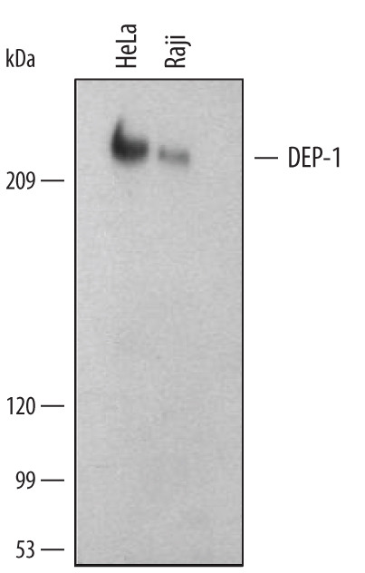
- Detection of Human DEP-1/CD148 by Western Blot. Western blot shows lysates of HeLa human cervical epithelial carcinoma cell line and Raji human Burkitt's lymphoma cell line. PVDF membrane was probed with 1 µg/mL of Human DEP-1/CD148 Monoclonal Antibody (Catalog # MAB1934) followed by HRP-conjugated Anti-Mouse IgG Secondary Antibody (Catalog # HAF007). A specific band was detected for DEP-1/CD148 at approximately 220 kDa (as indicated). This experiment was conducted under reducing conditions and using Immunoblot Buffer Group 1.