Explore
 Validate
 Learn
Learn Immunocytochemistry
Antibody data
- Antibody Data
- Antigen structure
- References [2]
- Comments [0]
- Validations
- Immunocytochemistry [2]
- Immunohistochemistry [5]
- Other assay [2]
Submit
Validation data
Reference
Comment
Report error
- Product number
- PA5-51665 - Provider product page

- Provider
- Invitrogen Antibodies
- Product name
- Fibulin 2 Polyclonal Antibody
- Antibody type
- Polyclonal
- Antigen
- Recombinant protein fragment
- Description
- Immunogen sequence: TLGSFHCYKA LTCEPGYALK DGECEDVDEC AMGTHTCQPG FLCQNTKGSF YCQARQRCMD GFLQDPEGNC VDINECTSLS EPCRPGFSCI NTVGSYTCQR NPLICARGYH ASDDGTKCVD VNE Highest antigen sequence identity to the following orthologs: Mouse - 85%, Rat - 85%.
- Reactivity
- Human
- Host
- Rabbit
- Isotype
- IgG
- Vial size
- 100 μL
- Concentration
- 0.2 mg/mL
- Storage
- Store at 4°C short term. For long term storage, store at -20°C, avoiding freeze/thaw cycles.
Submitted references Fibulin-2 expression associates with vascular invasion and patient survival in breast cancer.
Flow-induced Reorganization of Laminin-integrin Networks Within the Endothelial Basement Membrane Uncovered by Proteomics.
Klingen TA, Chen Y, Aas H, Wik E, Akslen LA
PloS one 2021;16(4):e0249767
PloS one 2021;16(4):e0249767
Flow-induced Reorganization of Laminin-integrin Networks Within the Endothelial Basement Membrane Uncovered by Proteomics.
Béguin EP, Janssen EFJ, Hoogenboezem M, Meijer AB, Hoogendijk AJ, van den Biggelaar M
Molecular & cellular proteomics : MCP 2020 Jul;19(7):1179-1192
Molecular & cellular proteomics : MCP 2020 Jul;19(7):1179-1192
No comments: Submit comment
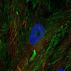
Supportive validation
- Submitted by
- Invitrogen Antibodies (provider)
- Main image

- Experimental details
- Immunofluorescent staining of Fibulin 2 in human cell line BJ using a Fibulin 2 Polyclonal Antibody (Product # PA5-51665) shows localization to plasma membrane.

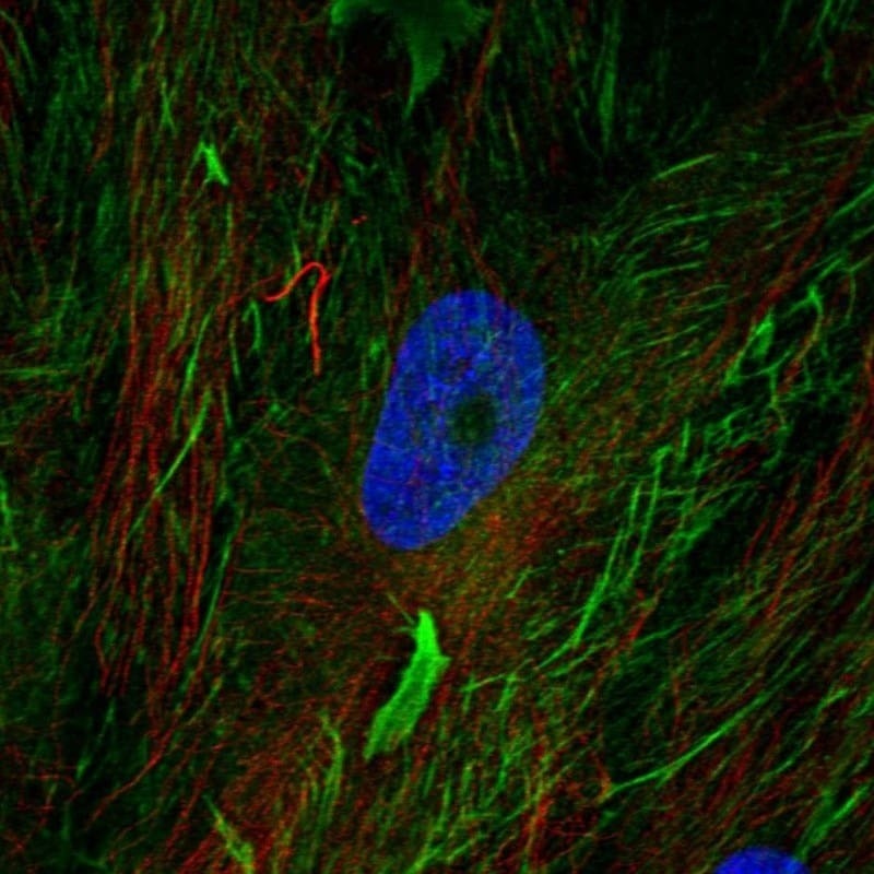
- Submitted by
- Invitrogen Antibodies (provider)
- Main image
- Experimental details
- Immunofluorescent staining of Fibulin 2 in human cell line BJ using a Fibulin 2 Polyclonal Antibody (Product # PA5-51665) shows localization to plasma membrane.
Supportive validation
- Submitted by
- Invitrogen Antibodies (provider)
- Main image
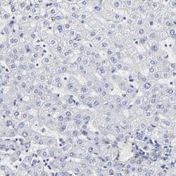
- Experimental details
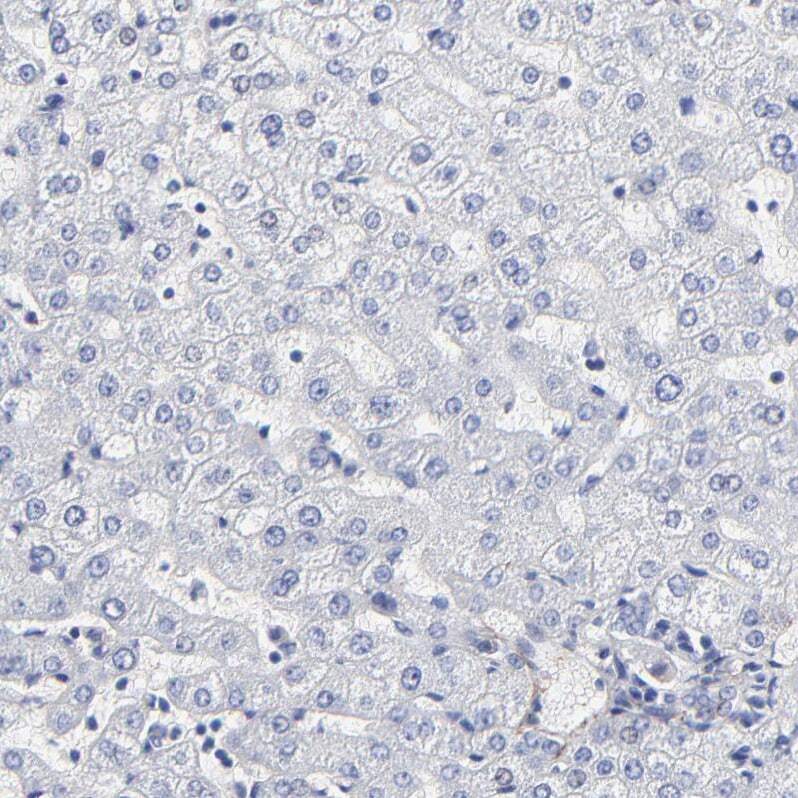
- Immunohistochemical analysis of Fibulin 2 in human liver using Fibulin 2 Polyclonal Antibody (Product # PA5-51665) shows no positivity in hepatocytes as expected.
- Submitted by
- Invitrogen Antibodies (provider)
- Main image
- Experimental details
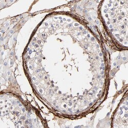
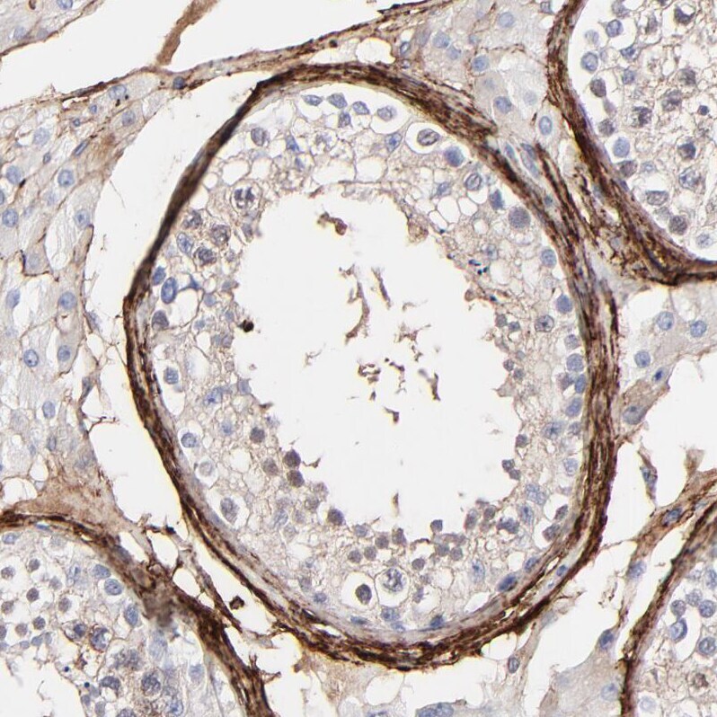
- Immunohistochemical analysis of Fibulin 2 in human testis using Fibulin 2 Polyclonal Antibody (Product # PA5-51665) shows strong membranous positivity in peritubular myoid cells.
- Submitted by
- Invitrogen Antibodies (provider)
- Main image
- Experimental details
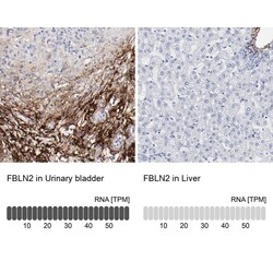
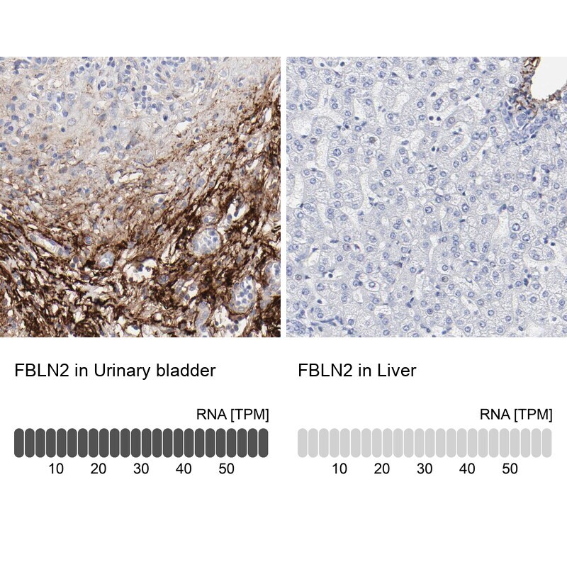
- Immunohistochemical staining of Fibulin 2 in human urinary bladder and liver tissues using Fibulin 2 Polyclonal Antibody (Product # PA5-51665). Corresponding FBLN2 RNA-seq data are presented for the same tissues.
- Submitted by
- Invitrogen Antibodies (provider)
- Main image
- Experimental details
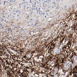
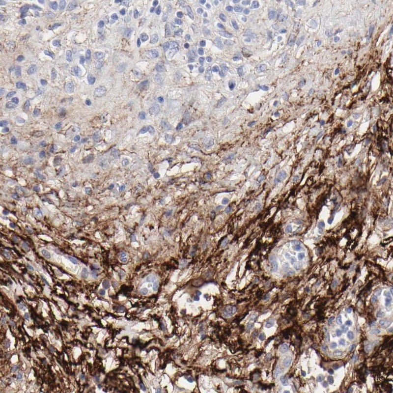
- Immunohistochemical staining of Fibulin 2 in human urinary bladder using Fibulin 2 Polyclonal Antibody (Product # PA5-51665) shows high expression.
- Submitted by
- Invitrogen Antibodies (provider)
- Main image

- Experimental details
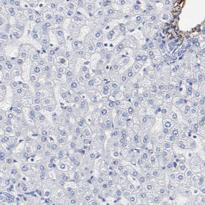
- Immunohistochemical staining of Fibulin 2 in human liver using Fibulin 2 Polyclonal Antibody (Product # PA5-51665) shows low expression as expected.
Supportive validation
- Submitted by
- Invitrogen Antibodies (provider)
- Main image
- Experimental details
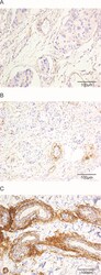
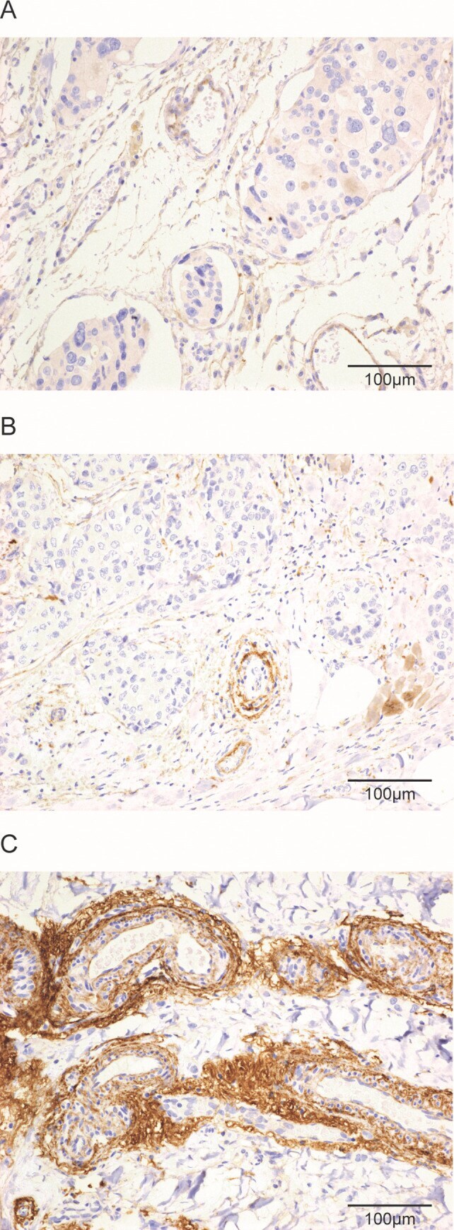
- Fig 1 Histological images of perivascular fibulin-2 expression by immuno-histochemistry. Histological images of tumour tissue with absent or faint/barely perceptible peripheral staining in tumour vessels (A), weak to moderate staining (B), or strong/thick staining (C) for fibulin-2 in vessels at the tumour periphery (x200). Scale bars are shown on each image.
- Submitted by
- Invitrogen Antibodies (provider)
- Main image

- Experimental details
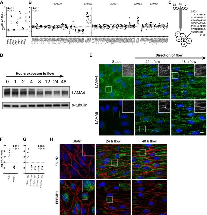
- Fig. 3. Flow induces remodeling of the endothelial extracellular matrix. A , Proteome and ( B ) cell surface proteome data of identified laminins. T or CT represents the enzyme used for protein digestion. The depicted numbers represent the biotin-modified lysine residue. C , Schematic representation of laminin 411 with the linker region between domains LG3 and LG4. The blue lysine residues were found to be decreased under flow conditions (as depicted in panel B ). D , Immunoblot analysis of LAMA4 in ECs exposed to flow over time. alpha-tubulin was used as a loading control. E , Protein distribution of LAMA5 and LAMA4 after flow-exposure. Green represents LAMA4 or LAMA5, blue is HOECHST. The scale bar is 10 mum. Depicted are maximum intensity projections. F , Proteome and ( G ) cell surface proteome data of identified fibulins. H , Immunofluorescence analysis of EFEMP1 and FBLN2 abundance and distribution affected by flow. Red is VE-cadherin, blue is HOECHST. Depicted are maximum intensity projections. Micrographs are representative for 3 independent experiments. The scale bar is 10 mum.