Explore
 Validate
 Learn
Learn Western blot
 Immunohistochemistry
Antibody data
- Antibody Data
- Antigen structure
- References [1]
- Comments [0]
- Validations
- Immunohistochemistry [1]
- Other assay [2]
Submit
Validation data
Reference
Comment
Report error
- Product number
- PA5-21640 - Provider product page

- Provider
- Invitrogen Antibodies
- Product name
- Fibulin 2 Polyclonal Antibody
- Antibody type
- Polyclonal
- Antigen
- Recombinant full-length protein
- Description
- Recommended positive controls: 293T, A431, HeLa, HepG2. Predicted reactivity: Mouse (86%). Store product as a concentrated solution. Centrifuge briefly prior to opening the vial.
- Reactivity
- Human, Mouse, Rat
- Host
- Rabbit
- Isotype
- IgG
- Vial size
- 100 μL
- Concentration
- 1.19 mg/mL
- Storage
- Store at 4°C short term. For long term storage, store at -20°C, avoiding freeze/thaw cycles.
Submitted references An Investigation of Fibulin-2 in Hypertrophic Cardiomyopathy.
Ibrahim AM, Roshdy M, Elshorbagy S, Hosny M, Halawa S, Yehia D, Elfawy HA, Eldessouki A, Mohamed F, Ellithy A, Abdelfattah M, Elsawy A, Elkhatib M, Allouba M, Elguindy A, Aguib Y, Yacoub M
International journal of molecular sciences 2020 Sep 29;21(19)
International journal of molecular sciences 2020 Sep 29;21(19)
No comments: Submit comment
Supportive validation
- Submitted by
- Invitrogen Antibodies (provider)
- Main image
- Experimental details
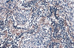
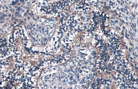
- Fibulin 2 Polyclonal Antibody detects Fibulin 2 protein at cell membrane by immunohistochemical analysis. Sample: Paraffin-embedded human endometrial carcinoma. Fibulin 2 stained by Fibulin 2 Polyclonal Antibody (Product # PA5-21640) diluted at 1:500. Antigen Retrieval: Citrate buffer, pH 6.0, 15 min.
Supportive validation
- Submitted by
- Invitrogen Antibodies (provider)
- Main image
- Experimental details
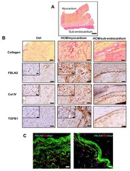
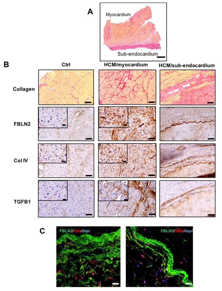
- Figure 2 FBLN2 expression and localization in HCM myectomy tissues along with Col IV and TGFbeta1. ( A ) A representative transmural section of a myectomy specimen stained with picro-Sirius red and shows fibrosis distribution in the myocardium and sub-endocardium. Scale bar is 1 mm. ( B ) Picro-sirius red and immunohistochemical analysis of HCM tissues at the myocardium and sub-endocardial ( n = 79), compared to controls ( n = 9), showing FBLN2, Col IV, and TGFbeta1 expression. Scale bars are 100 (overview) and 20 um (magnified insets). ( C ) Confocal microscopy imaging shows Vim+ and SMA+ cells (red) infiltrating the FBLN2 sheath (green) at the sub-endocardial region (representative of 3 HCMs vs. 3 controls). Scale bars are 20 um.
- Submitted by
- Invitrogen Antibodies (provider)
- Main image
- Experimental details
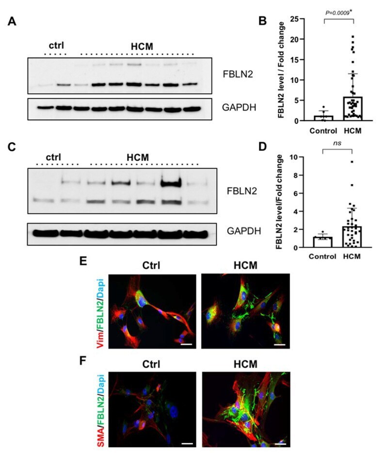
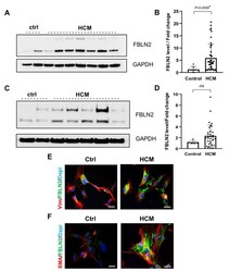
- Figure 3 Elevated expression of FBLN2 in HCM myectomy tissues and cultured fibroblasts. ( A ) A representative Immunoblot for HCM and ctrl tissues shows total FBLN2 protein expression normalized to Glyceraldehyde 3-phosphate dehydrogenase (GAPDH) expression. ( B ) Bar plot shows a significant difference ( P = 0.0009) between mean FBLN2 expression in HCM tissues ( n = 44) compared to ctrl tissues ( n = 7). ( C ) A representative Immunoblot for cultured HCM and ctrl fibroblasts shows total FBLN2 protein expression normalized to GAPDH expression. ( D ) Bar plot shows an elevated level of FBLN2 in cultured HCM fibroblasts ( n = 34) compared to ctrl fibroblasts ( n = 5). E and F: Confocal microscope imaging of cultured HCM ( n = 3) and control fibroblasts ( n = 3), shows FBLN2 expression pattern and localization. Cells were co-stained with FBLN2 and vimentin (mesenchymal fibroblasts marker) ( E ), and FBLN2 with smooth muscle actin (myofibroblasts marker) ( F ). Scale bars are 20 um.