 Explore
 Validate
 Learn
Learn Immunocytochemistry
 Immunohistochemistry
Antibody data
- Antibody Data
- Antigen structure
- References [1]
- Comments [0]
- Validations
- Immunocytochemistry [1]
Submit
Validation data
Reference
Comment
Report error
- Product number
- HPA018842 - Provider product page

- Provider
- Atlas Antibodies
- Proper citation
- Atlas Antibodies Cat#HPA018842, RRID:AB_1852864
- Product name
- Anti-LIAS
- Antibody type
- Polyclonal
- Description
- Polyclonal Antibody against Human LIAS, Gene description: lipoic acid synthetase, Alternative Gene Names: LAS, Validated applications: ICC, IHC, Uniprot ID: O43766, Storage: Store at +4°C for short term storage. Long time storage is recommended at -20°C.
- Reactivity
- Human, Mouse
- Host
- Rabbit
- Conjugate
- Unconjugated
- Isotype
- IgG
- Vial size
- 100 µl
- Concentration
- 0.1 mg/ml
- Storage
- Store at +4°C for short term storage. Long time storage is recommended at -20°C.
- Handling
- The antibody solution should be gently mixed before use.
Submitted references NOTCH4ΔL12_16 sensitizes lung adenocarcinomas to EGFR-TKIs through transcriptional down-regulation of HES1
Zhang B, Dong S, Wang J, Huang T, Zhao P, Xu J, Liu D, Fu L, Wang L, Wang G, Zou C
Nature Communications 2023;14(1)
Nature Communications 2023;14(1)
No comments: Submit comment
Supportive validation
- Submitted by
- Atlas Antibodies (provider)
- Main image
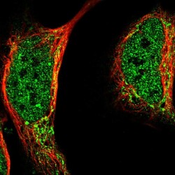
- Experimental details
- Immunofluorescent staining of human cell line U-2 OS shows localization to nucleoplasm & mitochondria.
- Sample type
- Human
