 Explore
 Validate
 Learn
Learn Western blot
Antibody data
- Antibody Data
- Antigen structure
- References [1]
- Comments [0]
- Validations
- Western blot [2]
- Immunocytochemistry [1]
Submit
Validation data
Reference
Comment
Report error
- Product number
- PA5-72230 - Provider product page

- Provider
- Invitrogen Antibodies
- Product name
- SOX17 Polyclonal Antibody
- Antibody type
- Polyclonal
- Antigen
- Synthetic peptide
- Reactivity
- Human, Mouse
- Host
- Rabbit
- Isotype
- IgG
- Vial size
- 400 µL
- Concentration
- 0.5 mg/mL
- Storage
- Store at 4°C short term. For long term storage, store at -20°C, avoiding freeze/thaw cycles.
Submitted references mRNA and miRNA expression profiles in an ectoderm-biased substate of human pluripotent stem cells.
Mawaribuchi S, Aiki Y, Ikeda N, Ito Y
Scientific reports 2019 Aug 15;9(1):11910
Scientific reports 2019 Aug 15;9(1):11910
No comments: Submit comment
Supportive validation
- Submitted by
- Invitrogen Antibodies (provider)
- Main image

- Experimental details
- Western blot analysis of (Mouse) Sox17 in the following samples: Lane 1: F9 cell lysate, Lane 2: LNCap cell lysate, Lane 3: mouse testis lysate. 20 µg of protein per lane was used. Samples were first blocked with 5% NFDM/TBST, incubated with (Mouse) Sox17 polyclonal antibody (Product # PA5-72230) using a dilution of 1:2000 followed by a goat anti-rabbit IgG H&L (HRP) secondary antibody with a dilution of 1:10,000.

- Submitted by
- Invitrogen Antibodies (provider)
- Main image
- Experimental details
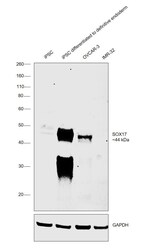
- Western Blot was performed using Anti-SOX17 Polyclonal Antibody (Product # PA5-72230) and a 44 kDa band corresponding to Transcription factor SOX-17 was observed in iPSC differentiated to definitive endoderm and OVCAR-3 when compared to other IMR-32. Nuclear enriched extracts (30 µg lysate) of iPSC (Lane 1), iPSC differentiated to definitive endoderm (Lane 2), OVCAR-3 (Lane 3), IMR-32 (Lane 4) were electrophoresed using NuPAGE™ 10% Bis-Tris Protein Gel (Product # NP0301BOX). Resolved proteins were then transferred onto a nitrocellulose membrane (Product # IB23001) by iBlot® 2 Dry Blotting System (Product # IB21001). The blot was probed with the primary antibody (1:2000 dilution) and detected by chemiluminescence with Goat anti-Rabbit IgG (H+L) Superclonal™ Recombinant Secondary Antibody, HRP (Product # A27036, 1:6000 dilution) using the iBright FL 1000 (Product # A32752). Chemiluminescent detection was performed using Novex® ECL Chemiluminescent Substrate Reagent Kit (Product # WP20005).

Supportive validation
- Submitted by
- Invitrogen Antibodies (provider)
- Main image
- Experimental details
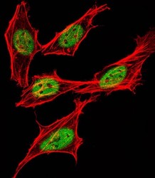
- Immunofluorescent analysis of Sox17 in HeLa cells. Samples were first fixed with 4% paraformaldehyde and permeabilized wit 0.1% Triton X-100. Cells were incubated with Sox17 polyclonal antibody (Product # PA5-72230) using a dilution of 1:25. Alexa Fluor 488-conjugated goat anti-rabbit lgG (green) with a dilution of 1:400 was used as the secondary antibody. Cytoplasmic actin was detected with Alexa Fluor 555 conjugated with Phalloidin at 1:100 dilution (red).
