 Explore
 Validate
 Learn
Learn Western blot
 Immunocytochemistry
 Flow cytometry
Antibody data
- Antibody Data
- Antigen structure
- References [1]
- Comments [0]
- Validations
- Immunocytochemistry [5]
Submit
Validation data
Reference
Comment
Report error
- Product number
- MA5-24885 - Provider product page

- Provider
- Invitrogen Antibodies
- Product name
- SOX17 Monoclonal Antibody (OTI3B10)
- Antibody type
- Monoclonal
- Antigen
- Recombinant protein fragment
- Reactivity
- Human
- Host
- Mouse
- Isotype
- IgG
- Antibody clone number
- OTI3B10
- Vial size
- 100 µL
- Concentration
- 1 mg/mL
- Storage
- -20° C, Avoid Freeze/Thaw Cycles
Submitted references Single-cell atlas unveils cellular heterogeneity and novel markers in human neonatal and adult intervertebral discs.
Jiang W, Glaeser JD, Salehi K, Kaneda G, Mathkar P, Wagner A, Ho R, Sheyn D
iScience 2022 Jul 15;25(7):104504
iScience 2022 Jul 15;25(7):104504
No comments: Submit comment
Supportive validation
- Submitted by
- Invitrogen Antibodies (provider)
- Main image
- Experimental details
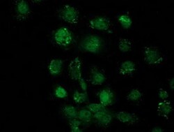
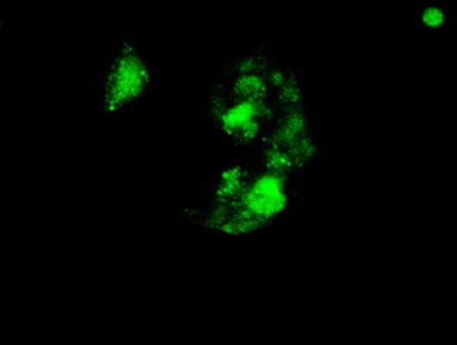
- Immunofluorescent analysis of SOX17 in KhES-3 cells. Cells were probed with a IdU monoclonal antibody (Product # MA5-24885).
- Submitted by
- Invitrogen Antibodies (provider)
- Main image
- Experimental details
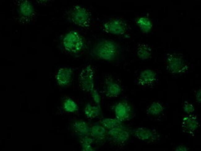
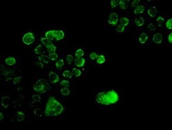
- Immunofluorescent analysis of SOX17 in KhES-3 cells. Cells were probed with a IdU monoclonal antibody (Product # MA5-24885).
- Submitted by
- Invitrogen Antibodies (provider)
- Main image

- Experimental details
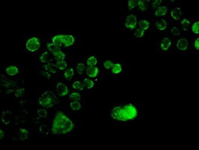
- Immunofluorescent analysis of SOX17 in KhES-3 cells. Cells were probed with a IdU monoclonal antibody (Product # MA5-24885).
- Submitted by
- Invitrogen Antibodies (provider)
- Main image
- Experimental details
- Immunofluorescent analysis of SOX17 in KhES-3 cells. Cells were probed with a IdU monoclonal antibody (Product # MA5-24885).
- Submitted by
- Invitrogen Antibodies (provider)
- Main image
- Experimental details
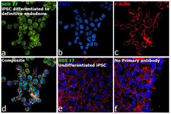
- Immunofluorescence analysis of Transcription factor SOX-17 was performed using 70% confluent log phase iPSC and iPSC differentiated to definitive endoderm cells. The cells were fixed with 4% paraformaldehyde for 10 minutes, permeabilized with 0.1% Triton™ X-100 for 15 minutes, and blocked with 2% BSA for 45 minutes at room temperature. The cells were labeled with SOX17 Monoclonal Antibody (OTI3B10) (Product # MA5-24885) at 1:100 dilution in 0.1% BSA, incubated at 4 degree celsius overnight and then labeled with Donkey anti-Mouse IgG (H+L) Highly Cross-Adsorbed Secondary Antibody, Alexa Fluor Plus 488 (Product # A32766), (1:2000 dilution), for 45 minutes at room temperature (Panel a: Green). Nuclei (Panel b: Blue) were stained with ProLong™ Diamond Antifade Mountant with DAPI (Product # P36962). F-actin (Panel c: Red) was stained with Rhodamine Phalloidin (Product # R415, 1:300). Panel d represents the merged image showing nuclear localization. Panel e represents control iPSC which shows no signal. Panel f represents control cells with no primary antibody to assess background. The images were captured at 60X magnification.
