Explore
 Validate
 Learn
Learn Western blot
 Immunocytochemistry
Antibody data
- Antibody Data
- Antigen structure
- References [1]
- Comments [0]
- Validations
- Western blot [6]
- Immunohistochemistry [1]
Submit
Validation data
Reference
Comment
Report error
- Product number
- NBP2-17086 - Provider product page

- Provider
- Novus Biologicals
- Product name
- Rabbit Polyclonal KMT1A/SUV39H1 Antibody
- Antibody type
- Polyclonal
- Description
- Immunogen affinity purified.
- Reactivity
- Human, Mouse, Simian
- Host
- Rabbit
- Isotype
- IgG
- Vial size
- 0.1 ml
- Storage
- Aliquot and store at -20C or -80C. Avoid freeze-thaw cycles.
Submitted references Super-resolution imaging reveals the evolution of higher-order chromatin folding in early carcinogenesis.
Xu J, Ma H, Ma H, Jiang W, Mela CA, Duan M, Zhao S, Gao C, Hahm ER, Lardo SM, Troy K, Sun M, Pai R, Stolz DB, Zhang L, Singh S, Brand RE, Hartman DJ, Hu J, Hainer SJ, Liu Y
Nature communications 2020 Apr 20;11(1):1899
Nature communications 2020 Apr 20;11(1):1899
No comments: Submit comment
Supportive validation
- Submitted by
- Novus Biologicals (provider)
- Main image
- Experimental details
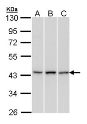
- Western Blot: KMT1A/SUV39H1 Antibody [NBP2-17086] - Sample (30 ug of whole cell lysate) A: H1299 B: Hep G2 C: Raji 10% SDS PAGE gel, diluted at 1:1000.

- Submitted by
- Novus Biologicals (provider)
- Main image

- Experimental details
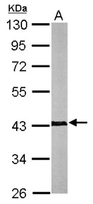
- Western Blot: KMT1A/SUV39H1 Antibody [NBP2-17086] - Sample (50 ug of whole cell lysate) A: Mouse Brain, 10% SDS PAGE gel, diluted at 1:1000.
- Submitted by
- Novus Biologicals (provider)
- Main image
- Experimental details
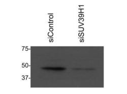
- Western Blot: KMT1A/SUV39H1 Antibody [NBP2-17086] - Western blot with anti Suv39H1 after siControl and siSUV39H1 in MCF7 cells. Image from verified customer review.

- Submitted by
- Novus Biologicals (provider)
- Main image
- Experimental details
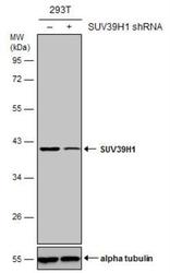
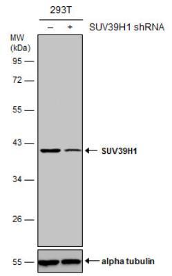
- Western Blot: KMT1A/SUV39H1 Antibody [NBP2-17086] - Non-transfected (-) and transfected (+) 293T whole cell extracts (50 ug) were separated by 10% SDS-PAGE, and the membrane was blotted with SUV39H1 antibody [N3C3]. HRP-conjugated anti-rabbit IgG antibody was used to detect the primary antibody.
- Submitted by
- Novus Biologicals (provider)
- Main image
- Experimental details
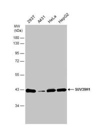
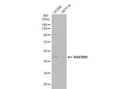
- Western Blot: KMT1A/SUV39H1 Antibody [NBP2-17086] - Various whole cell extracts (30 ug) were separated by 10% SDS-PAGE, and the membrane was blotted with SUV39H1 antibody [N3C3] diluted at 1:1000.
- Submitted by
- Novus Biologicals (provider)
- Main image
- Experimental details
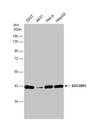
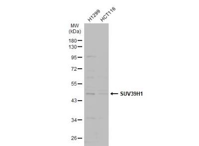
- Western Blot: KMT1A/SUV39H1 Antibody [NBP2-17086] - Various whole cell extracts (30 ug) were separated by 10% SDS-PAGE, and the membrane was blotted with SUV39H1 antibody [N3C3] diluted at 1:500. The HRP-conjugated anti-rabbit IgG antibody (NBP2-19301) was used to detect the primary antibody.
Supportive validation
- Submitted by
- Novus Biologicals (provider)
- Main image
- Experimental details
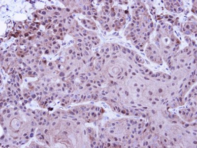
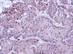
- Immunohistochemistry-Paraffin: KMT1A/SUV39H1 Antibody [NBP2-17086] - Immunohistochemical analysis of paraffin-embedded Cal27 Xenograft, using antibody at 1:100 dilution.