Explore
 Validate
 Learn
Learn Western blot
 Immunoprecipitation
Antibody data
- Antibody Data
- Antigen structure
- References [5]
- Comments [0]
- Validations
- Western blot [6]
- Immunocytochemistry [1]
- Immunohistochemistry [1]
Submit
Validation data
Reference
Comment
Report error
- Product number
- GTX112263 - Provider product page

- Provider
- GeneTex
- Proper citation
- GeneTex Cat#GTX112263, RRID:AB_1952113
- Product name
- SUV39H1 antibody [N3C3]
- Antibody type
- Polyclonal
- Reactivity
- Human, Mouse, Simian
- Host
- Rabbit
Submitted references G9a governs colon cancer stem cell phenotype and chemoradioresistance through PP2A-RPA axis-mediated DNA damage response.
Inhibition of KDM4A activity as a strategy to suppress interleukin-6 production and attenuate colitis induction.
In vitro hydroquinone-induced instauration of histone bivalent mark on human retroelements (LINE-1) in HL60 cells.
Role of the histone H3 lysine 9 methyltransferase Suv39 h1 in maintaining Epsteinn-Barr virus latency in B95-8 cells.
The SRA protein UHRF1 promotes epigenetic crosstalks and is involved in prostate cancer progression.
Luo CW, Wang JY, Hung WC, Peng G, Tsai YL, Chang TM, Chai CY, Lin CH, Pan MR
Radiotherapy and oncology : journal of the European Society for Therapeutic Radiology and Oncology 2017 Sep;124(3):395-402
Radiotherapy and oncology : journal of the European Society for Therapeutic Radiology and Oncology 2017 Sep;124(3):395-402
Inhibition of KDM4A activity as a strategy to suppress interleukin-6 production and attenuate colitis induction.
Ishiguro K, Watanabe O, Nakamura M, Yamamura T, Matsushita M, Goto H, Hirooka Y
Clinical immunology (Orlando, Fla.) 2017 Jul;180:120-127
Clinical immunology (Orlando, Fla.) 2017 Jul;180:120-127
In vitro hydroquinone-induced instauration of histone bivalent mark on human retroelements (LINE-1) in HL60 cells.
Mancini M, Mandruzzato M, Garzia AC, Sahnane N, Magnani E, Macchi F, Oulad-Abdelghani M, Oudet P, Bollati V, Fustinoni S, Furlan D, Bonapace IM
Toxicology in vitro : an international journal published in association with BIBRA 2017 Apr;40:1-10
Toxicology in vitro : an international journal published in association with BIBRA 2017 Apr;40:1-10
Role of the histone H3 lysine 9 methyltransferase Suv39 h1 in maintaining Epsteinn-Barr virus latency in B95-8 cells.
Imai K, Kamio N, Cueno ME, Saito Y, Inoue H, Saito I, Ochiai K
The FEBS journal 2014 May;281(9):2148-58
The FEBS journal 2014 May;281(9):2148-58
The SRA protein UHRF1 promotes epigenetic crosstalks and is involved in prostate cancer progression.
Babbio F, Pistore C, Curti L, Castiglioni I, Kunderfranco P, Brino L, Oudet P, Seiler R, Thalman GN, Roggero E, Sarti M, Pinton S, Mello-Grand M, Chiorino G, Catapano CV, Carbone GM, Bonapace IM
Oncogene 2012 Nov 15;31(46):4878-87
Oncogene 2012 Nov 15;31(46):4878-87
No comments: Submit comment
Supportive validation
- Submitted by
- GeneTex (provider)
- Main image

- Experimental details
- Sample (30 ?g of whole cell lysate) A: H1299 B: HepG2 (GTX27900) C: Raji 10% SDS PAGE GTX112263 diluted at 1:1000 The HRP-conjugated anti-rabbit IgG antibody (GTX213110-01) was used to detect the primary antibody.
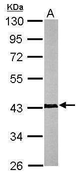
- Submitted by
- GeneTex (provider)
- Main image

- Experimental details
- Sample (50 ?g of whole cell lysate) A: mouse brain 10% SDS PAGE GTX112263 diluted at 1:1000 The HRP-conjugated anti-rabbit IgG antibody (GTX213110-01) was used to detect the primary antibody.
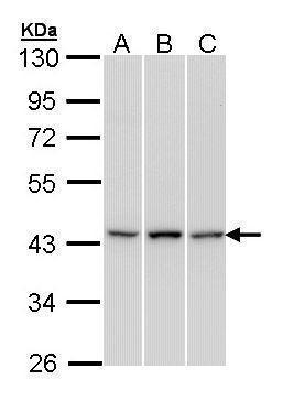
- Submitted by
- GeneTex (provider)
- Main image
- Experimental details
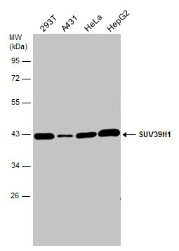
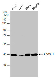
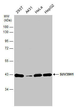
- Various whole cell extracts (30 ?g) were separated by 10% SDS-PAGE, and the membrane was blotted with SUV39H1 antibody [N3C3] (GTX112263) diluted at 1:1000. The HRP-conjugated anti-rabbit IgG antibody (GTX213110-01) was used to detect the primary antibody.
- Submitted by
- GeneTex (provider)
- Main image
- Experimental details
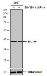
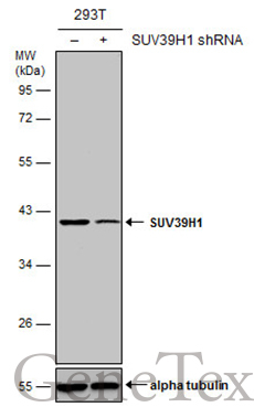
- Non-transfected (¡V) and transfected (+) 293T whole cell extracts (50 ?g) were separated by 10% SDS-PAGE, and the membrane was blotted with SUV39H1 antibody [N3C3] (GTX112263) diluted at 1:10000. The HRP-conjugated anti-rabbit IgG antibody (GTX213110-01) was used to detect the primary antibody.
- Submitted by
- GeneTex (provider)
- Main image

- Experimental details
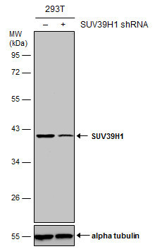
- Non-transfected (¡V) and transfected (+) 293T whole cell extracts (50 ?g) were separated by 10% SDS-PAGE, and the membrane was blotted with SUV39H1 antibody [N3C3] (GTX112263) diluted at 1:10000. The HRP-conjugated anti-rabbit IgG antibody (GTX213110-01) was used to detect the primary antibody.
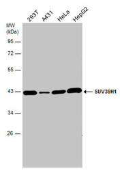
- Submitted by
- GeneTex (provider)
- Main image
- Experimental details
- Various whole cell extracts (30 ?g) were separated by 10% SDS-PAGE, and the membrane was blotted with SUV39H1 antibody [N3C3] (GTX112263) diluted at 1:1000.
Supportive validation
- Submitted by
- GeneTex (provider)
- Main image
- Experimental details
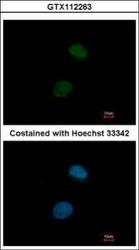
- Immunofluorescence analysis of paraformaldehyde-fixed HeLa, using SUV39H1(GTX112263) antibody at 1:200 dilution.

Supportive validation
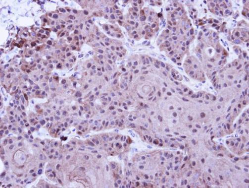
- Submitted by
- GeneTex (provider)
- Main image

- Experimental details
- Immunohistochemical analysis of paraffin-embedded Cal27 Xenograft, using SUV39H1(GTX112263) antibody at 1:100 dilution.