Explore
 Validate
 Learn
Learn Western blot
 Immunocytochemistry
 Immunoprecipitation
Antibody data
- Antibody Data
- Antigen structure
- References [1]
- Comments [0]
- Validations
- Immunocytochemistry [5]
- Immunohistochemistry [1]
- Other assay [1]
Submit
Validation data
Reference
Comment
Report error
- Product number
- PA5-29470 - Provider product page

- Provider
- Invitrogen Antibodies
- Product name
- SUV39H1 Polyclonal Antibody
- Antibody type
- Polyclonal
- Antigen
- Recombinant full-length protein
- Description
- Recommended positive controls: 293T, A431, HeLa, HepG2, H1299, Raji, Mouse brain. Predicted reactivity: Mouse (98%), Rat (98%), Xenopus laevis (90%), Rhesus Monkey (100%), Bovine (100%). Store product as a concentrated solution. Centrifuge briefly prior to opening the vial.
- Reactivity
- Human, Mouse
- Host
- Rabbit
- Isotype
- IgG
- Vial size
- 100 μL
- Concentration
- 0.52 mg/mL
- Storage
- Store at 4°C short term. For long term storage, store at -20°C, avoiding freeze/thaw cycles.
Submitted references Heterochromatin-Driven Nuclear Softening Protects the Genome against Mechanical Stress-Induced Damage.
Nava MM, Miroshnikova YA, Biggs LC, Whitefield DB, Metge F, Boucas J, Vihinen H, Jokitalo E, Li X, García Arcos JM, Hoffmann B, Merkel R, Niessen CM, Dahl KN, Wickström SA
Cell 2020 May 14;181(4):800-817.e22
Cell 2020 May 14;181(4):800-817.e22
No comments: Submit comment
Supportive validation
- Submitted by
- Invitrogen Antibodies (provider)
- Main image
- Experimental details
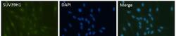
- Immunofluorescent analysis of SUV39H1 in paraformaldehyde-fixed HeLa cells using a SUV39H1 polyclonal antibody (Product # PA5-29470) at a 1:200 dilution.
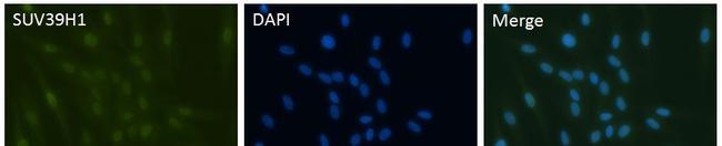
- Submitted by
- Invitrogen Antibodies (provider)
- Main image
- Experimental details
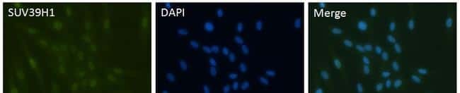
- Immunofluorescent analysis of SUV39H1 (green) in human hepatocytes. The cells were fixed, permeabilized and blocked. Cells were stained with a SUV39H1 polyclonal antibody (Product # PA5-29470) at a dilution of 1:500, followed by staining with Alexa fluor 488 goat anti-rabbit secondary antibody (Product # A-11008) at a dilution of 1:500 (green). Nuclei (blue) were counterstained with DAPI. Images were taken at 50X magnification. Data courtesy of the Antibody Data Exchange Program.
- Submitted by
- Invitrogen Antibodies (provider)
- Main image
- Experimental details
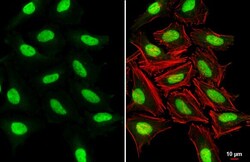
- SUV39H1 Polyclonal Antibody detects SUV39H1 protein at nucleus by immunofluorescent analysis. Sample: HeLa cells were fixed in 4% paraformaldehyde at RT for 15 min. Green: SUV39H1 stained by SUV39H1 Polyclonal Antibody (Product # PA5-29470) diluted at 1:500. Red: phalloidin, a cytoskeleton marker, diluted at 1:200. Scale bar= 10 µm.
- Submitted by
- Invitrogen Antibodies (provider)
- Main image

- Experimental details
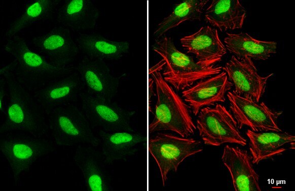
- SUV39H1 Polyclonal Antibody detects SUV39H1 protein at nucleus by immunofluorescent analysis. Sample: HeLa cells were fixed in 4% paraformaldehyde at RT for 15 min. Green: SUV39H1 stained by SUV39H1 Polyclonal Antibody (Product # PA5-29470) diluted at 1:500. Red: phalloidin, a cytoskeleton marker, diluted at 1:200. Scale bar= 10 µm.

- Submitted by
- Invitrogen Antibodies (provider)
- Main image
- Experimental details
- Immunofluorescent analysis of SUV39H1 (green) in human hepatocytes. The cells were fixed, permeabilized and blocked. Cells were stained with a SUV39H1 polyclonal antibody (Product # PA5-29470) at a dilution of 1:500, followed by staining with Alexa fluor 488 goat anti-rabbit secondary antibody (Product # A-11008) at a dilution of 1:500 (green). Nuclei (blue) were counterstained with DAPI. Images were taken at 50X magnification. Data courtesy of the Antibody Data Exchange Program.
Supportive validation
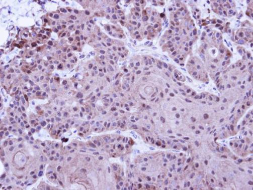
- Submitted by
- Invitrogen Antibodies (provider)
- Main image

- Experimental details
- Immunohistochemical analysis of paraffin-embedded Cal27 Xenograft, using SUV39H1 (Product # PA5-29470) antibody at 1:100 dilution. Antigen Retrieval: EDTA based buffer, pH 8.0, 15 min.
Supportive validation
- Submitted by
- Invitrogen Antibodies (provider)
- Main image

- Experimental details
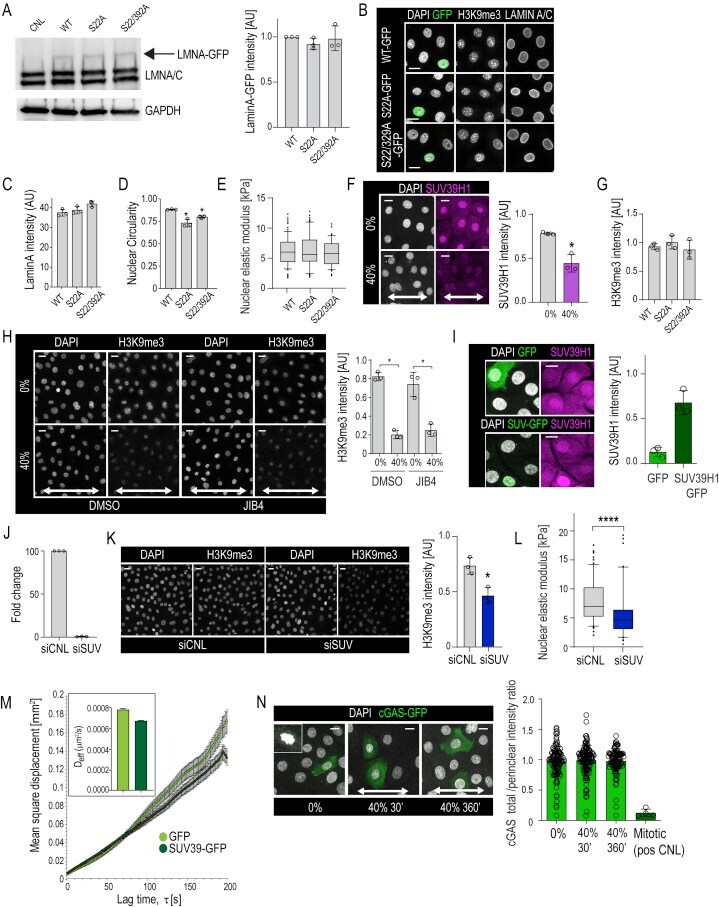
- Figure S4 H3K9me3 Regulates Chromatin Rheology and Nuclear Mechanics, Related to Figure 4 (A) Representative western blots and quantification of GFP-tagged wild-type (WT) Lamin A or Lamin A mutants (Serine 22 to Alanine (S22A) or S22A/S392A double mutant) show equal levels of expression. Lamin A band intensities are normalized to GAPDH (n = 3 independent experiments). (B) Representative immunofluorescence images of nuclei (DAPI), GFP, Lamin A/C, and H3K9me3 in cells expressing WT Lamin A-GFP or Lamin mutants S22A-GFP; S22A/S392A-GFP. (C and D) Quantification of Lamin A intensity and nuclear shape in WT Lamin A and mutant expressing cells (n = 3 independent experiments with > 100 nuclei/condition/experiment; * p = 0.034, ANOVA/Dunnett's). (E) AFM force indentation experiments of cells expressing WT Lamin A-GFP or Lamin mutants S22A-GFP; S22A/S392A-GFP (n = 3 independent experiments with > 40 nuclei/condition/experiment; no statistically significant difference found, Friedman/Dunn's). (F) Representative immunofluorescence images and quantification of Suv39H1 levels in cells exposed to uniaxial 40% stretch for 30 min (n = 3 independent experiments with > 100 nuclei/condition/experiment; * p = 0.05, Mann-Whitney) (G) Quantification of H3K9me3 intensity from cells in (B) (n = 3 independent experiments with > 30 nuclei/condition/experiment; no statistically significant difference found, Friedman/Dunn's). (H) Representative immunofluorescence images and quantification of H3K9me3 lev